 Back
Blood: Structure, Function, and Clinical Aspects
Study Guide - Smart Notes
 Tailored notes based on your materials, expanded with key definitions, examples, and context.
Blood: Structure, Function, and Clinical Aspects
Introduction to Blood
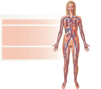
Blood is a specialized connective tissue that serves as the internal transport system of the body. It is essential for the distribution of nutrients, gases, hormones, and waste products, as well as for defense and temperature regulation.
Functions: Transport, regulation, and protection.
Volume: Approximately 5 liters in an average adult.
Properties: Slightly alkaline (pH ~7.4), temperature around 38°C, and about five times more viscous than water.
Composition of Blood
Plasma and Formed Elements
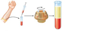
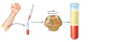
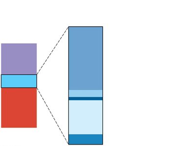
Blood consists of a liquid matrix called plasma and cellular components known as formed elements.
Plasma: Makes up about 55% of blood volume; contains water, proteins (albumins, globulins, fibrinogen), nutrients, hormones, and waste products.
Formed Elements: Include erythrocytes (red blood cells), leukocytes (white blood cells), and platelets (thrombocytes).
Formed Elements: Overview
The formed elements of blood are the cellular components responsible for transport, defense, and clotting.
Erythrocytes (RBCs): Transport oxygen and carbon dioxide.
Leukocytes (WBCs): Defend against pathogens.
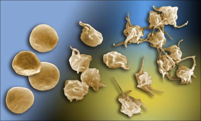
Platelets: Essential for blood clotting (hemostasis).
Erythrocytes (Red Blood Cells)
Structure and Function
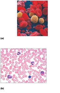
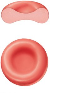
Erythrocytes are biconcave, anucleate cells specialized for gas transport. Their shape increases surface area for efficient gas exchange.
Diameter: ~7.5 μm
Lifespan: ~120 days
Main protein: Hemoglobin, which binds oxygen and carbon dioxide.
Hemoglobin
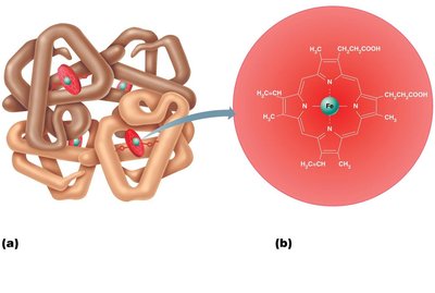
Hemoglobin is a complex protein composed of four polypeptide chains (two alpha, two beta) and four heme groups, each containing an iron ion that binds oxygen.
Oxygen transport: Each hemoglobin molecule can carry up to four oxygen molecules.
Carbon dioxide transport: Some CO2 binds to globin chains.
Erythropoiesis (Red Blood Cell Formation)
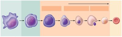
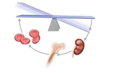
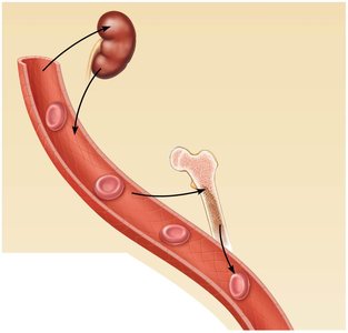
Erythropoiesis is the process of RBC production, occurring in the red bone marrow. It is regulated by the hormone erythropoietin (EPO), primarily produced by the kidneys in response to hypoxia (low oxygen levels).
Stages: Hematopoietic stem cell → Proerythroblast → Erythroblast stages → Reticulocyte → Erythrocyte
Regulation: EPO stimulates erythropoiesis when oxygen levels are low.
Red Blood Cell Life Cycle and Breakdown
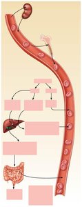
Old or damaged erythrocytes are removed by macrophages in the spleen, liver, and bone marrow. Hemoglobin is broken down into heme and globin; iron is recycled, and bilirubin is excreted in bile.
Iron recycling: Stored as ferritin or hemosiderin, transported by transferrin.
Bilirubin: Excreted in feces as stercobilin.
Disorders of Erythrocytes
Anemia: Reduced oxygen-carrying capacity of blood due to blood loss, decreased RBC production, or increased RBC destruction.
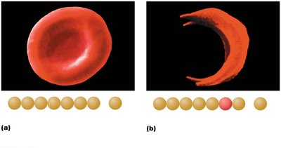
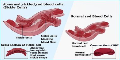
Sickle Cell Anemia: Genetic disorder causing abnormal hemoglobin, resulting in sickle-shaped RBCs that can block blood flow.
Polycythemia: Excess RBCs, increasing blood viscosity; can be caused by high altitude, EPO doping, or bone marrow disorders.

Leukocytes (White Blood Cells)
Types and Functions
Leukocytes are involved in immune defense and are classified as granulocytes or agranulocytes based on the presence of cytoplasmic granules.
Granulocytes: Neutrophils, eosinophils, basophils
Agranulocytes: Lymphocytes, monocytes
Leukocyte Formation (Leukopoiesis)
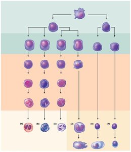
Leukocytes originate from hematopoietic stem cells in the bone marrow and differentiate into various lineages.
Myeloid lineage: Produces granulocytes and monocytes.
Lymphoid lineage: Produces lymphocytes (B and T cells).
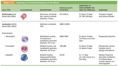
Summary Table: Formed Elements of Blood
Cell Type | Description | Cells/μL | Lifespan | Function |
|---|---|---|---|---|
Erythrocytes | Biconcave, anucleate | 4–6 million | 100–120 days | Transport O2 and CO2 |
Neutrophils | Multilobed nucleus | 3000–7000 | 6 hours–few days | Phagocytize bacteria |
Eosinophils | Bilobed nucleus, red granules | 100–400 | 5 days | Kill parasitic worms, allergy |
Basophils | Lobed nucleus, blue granules | 20–50 | Few hours–few days | Release histamine, anticoagulant |
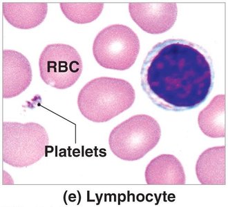
Lymphocytes | Large nucleus | 1500–3000 | Hours–years | Immune response |
Monocytes | Kidney-shaped nucleus | 100–700 | Months | Phagocytosis, macrophages |
Platelets | Cell fragments | 150,000–400,000 | 5–10 days | Clotting |
Leukocyte Disorders
Leukopenia: Abnormally low WBC count; increases risk of infection.
Leukemia: Cancer of WBCs; classified as acute or chronic, and by cell type (myeloid or lymphoid).
Platelets and Hemostasis
Platelet Formation
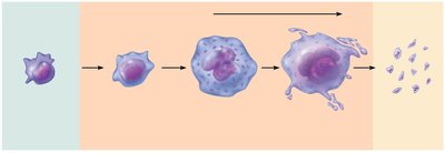
Platelets are fragments of megakaryocytes and are essential for blood clotting.
Hemostasis: Stopping Blood Loss
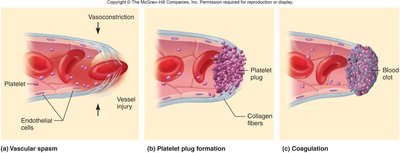
Hemostasis is the process that prevents blood loss after vessel injury. It involves three main steps:
Vascular spasm: Vasoconstriction reduces blood flow.
Platelet plug formation: Platelets adhere to exposed collagen and aggregate.
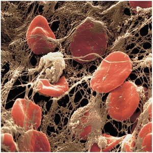
Coagulation: Fibrin forms a mesh that stabilizes the clot.
Clotting Disorders
Hemophilia: Genetic deficiency of clotting factors; leads to excessive bleeding.
Thrombocytopenia: Low platelet count; causes bleeding tendency.
Thrombosis: Formation of abnormal clots in unbroken vessels.
Embolus: Traveling clot that can block blood vessels.
Blood Types and Transfusion
ABO Blood Groups
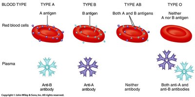
Blood types are determined by the presence or absence of antigens (A, B) on the surface of RBCs. Plasma contains antibodies against antigens not present on the individual's RBCs.
Rh Factor
The Rh antigen (D antigen) determines if blood type is positive or negative. Rh-negative individuals can develop antibodies if exposed to Rh-positive blood.
Transfusion Reactions
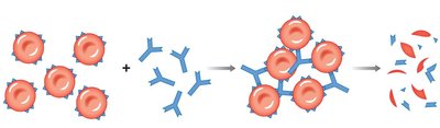
If incompatible blood is transfused, antibodies in the recipient's plasma react with donor RBC antigens, causing agglutination and hemolysis.
Hemolytic Disease of the Newborn (HDN)
Occurs when an Rh-negative mother is sensitized to Rh antigen and produces antibodies that attack the RBCs of an Rh-positive fetus in subsequent pregnancies.
Summary
Blood is vital for transport, defense, and homeostasis.
Its components include plasma, erythrocytes, leukocytes, and platelets.
Disorders can affect any component, leading to clinical consequences.
Blood typing and compatibility are crucial for safe transfusions.
