 Back
BackBlood: Structure, Function, and Clinical Relevance
Study Guide - Smart Notes
 Tailored notes based on your materials, expanded with key definitions, examples, and context.
Blood: Structure and Function
Overview of Blood as a Connective Tissue
Blood is a specialized connective tissue composed of cells (formed elements) suspended in a liquid matrix called plasma. It plays a vital role in transport, immunity, and homeostasis.
Cells: Red blood cells (RBCs), white blood cells (WBCs), and platelets
Matrix: Plasma (liquid component)
Definition of Connective Tissue: Characterized by scattered cells, matrix, ground substance, and fibers
Functions of Blood
Transport: Delivers oxygen to tissues and removes carbon dioxide
Immune Function: Mediated by WBCs and antibodies
Buffering: Maintains pH via the bicarbonate buffer system and carbonic anhydrase
Characteristics of Blood
Total volume: 5–6 L in adults
RBC count: 4–6 million/mm3
WBC count: 5,000–10,000/mm3
pH: 7.35–7.45 (slightly alkaline)
Viscosity: More viscous than water due to dissolved proteins
Hematocrit
Hematocrit is the percentage of RBCs in a blood sample. Normal range: 38–55% (higher in males).
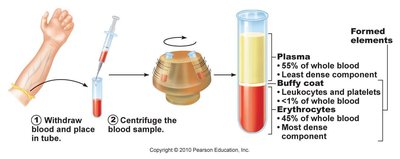
Plasma
Composition and Function
Plasma is a colloid, primarily water (~90%), containing dissolved substances such as carbohydrates, proteins, ions, and lipoproteins.
Proteins: Albumin (osmotic pressure, transport), globulins (immune function), fibrinogen (clotting)
Ions: K+, Na+, Cl-, HCO3-, Ca2+
Other solutes: Glucose, amino acids, lipids, hormones, waste products
Albumin
Maintains osmotic pressure, preventing edema
Produced in the liver
Transports substances such as bilirubin and fatty acids
Formed Elements
Red Blood Cells (Erythrocytes)
RBCs are anucleate cells specialized for oxygen transport. They are produced in the bone marrow and have a lifespan of ~120 days.
Hemoglobin: Protein with four globin chains, each containing a heme group with iron that binds oxygen
Normal hemoglobin: 13.5–17.5 g/dL (men), 12–15.5 g/dL (women)
Enzyme: Carbonic anhydrase (converts CO2 to bicarbonate)
Hematopoiesis and Erythropoiesis
Blood cells are formed in red bone marrow from pluripotent stem cells (hemocytoblasts). Erythropoiesis is the process of RBC formation, stimulated by erythropoietin (EPO) from the kidneys in response to hypoxia.
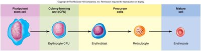
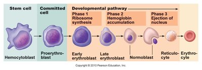
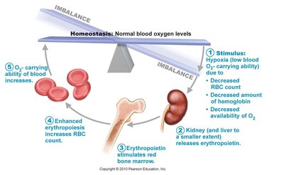
Reticulocyte: Last immature stage before mature RBC; increased reticulocytes indicate increased erythropoiesis
Fate of Red Blood Cells
Destroyed in the liver and spleen when aged
Hemoglobin breakdown: iron is recycled, globin is degraded to amino acids, heme is converted to bilirubin (excreted in bile)
Red Blood Cell Disorders
Polycythemia: Excess RBCs; increases blood viscosity
Anemia: Decreased oxygen-carrying capacity (various types: hemorrhagic, hemolytic, sickle cell, pernicious, aplastic)
Blood Typing and Transfusion
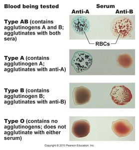
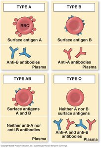
ABO Blood Group System
Blood types are determined by the presence of specific glycoprotein antigens (agglutinogens) on RBC surfaces. The main types are A, B, AB, and O.
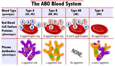
Type A: A antigen, anti-B antibodies
Type B: B antigen, anti-A antibodies
Type AB: Both A and B antigens, no antibodies (universal recipient)
Type O: No antigens, both anti-A and anti-B antibodies (universal donor)
Rh Factor and Hemolytic Disease of the Newborn
The Rh (D) antigen determines positive or negative blood type. Rh incompatibility can cause erythroblastosis fetalis in newborns if an Rh-negative mother carries an Rh-positive fetus.
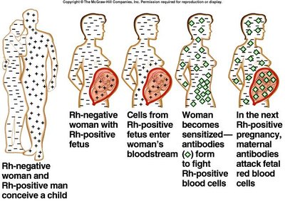
Prevention: Rh-negative mothers receive Rh immunoglobulin (Rhogam) to prevent antibody formation
White Blood Cells (Leukocytes)
General Characteristics
Contain nuclei and organelles
5,000–10,000/mm3 in blood; many reside in tissues
Functions: immune defense, diapedesis (migration through vessel walls), chemotaxis, phagocytosis
Types of White Blood Cells
Granulocytes: Neutrophils, eosinophils, basophils
Agranulocytes: Lymphocytes, monocytes
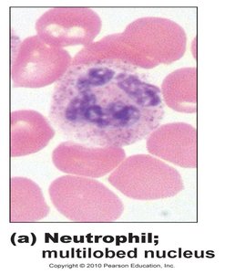
Neutrophils
Most abundant (50–70%)
First responders to infection; phagocytize bacteria
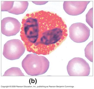
Eosinophils
2–5% of WBCs
Bilobed nucleus, reddish-orange granules
Combat parasitic infections and mediate allergic responses
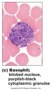
Basophils
0–1% of WBCs
Release histamine and heparin; mediate inflammation and allergic reactions
Monocytes
3–8% of WBCs
Phagocytic; become macrophages in tissues
Lymphocytes
20–40% of WBCs
T cells (cell-mediated immunity), B cells (antibody production), NK cells (immune surveillance)
WBC Disorders
Leukopenia: Low WBC count; increased infection risk
Leukocytosis: High WBC count; often due to infection or inflammation
Leukemia: Cancer of blood-forming tissues; abnormal proliferation of WBCs
Platelets and Hemostasis
Platelets (Thrombocytes)
Cell fragments derived from megakaryocytes
200,000–400,000/mm3
Essential for blood clotting (hemostasis)
Hemostasis: Stopping Bleeding
Vasoconstriction: Narrowing of blood vessels to reduce blood loss (mediated by serotonin)
Platelet Plug Formation: Platelets adhere to exposed collagen, aggregate, and release chemicals (ADP, serotonin, thromboxane A2)
Coagulation: Formation of a stable fibrin clot via the coagulation cascade
Clot Retraction: Fibrin strands contract, reducing clot size and aiding tissue repair
Coagulation Cascade
The coagulation cascade involves intrinsic and extrinsic pathways leading to the formation of fibrin, which stabilizes the clot.
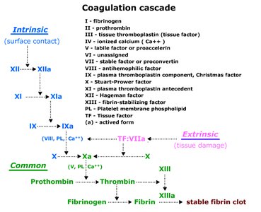
Key steps:
Formation of prothrombinase
Conversion of prothrombin to thrombin
Conversion of fibrinogen to fibrin
Clot Dissolution (Fibrinolysis)
Plasminogen is converted to plasmin, which digests fibrin and dissolves the clot
Clotting Disorders
Hemophilia: Genetic deficiency of clotting factors
Thrombosis: Abnormal clot formation in unbroken vessels
Embolus: A clot fragment traveling in the bloodstream
Key Vocabulary and Reference Values
RBC count: 4–6 million/mm3
Hemoglobin: 12–18 g/dL
Hematocrit: 38–55%
Total blood volume: 4–6 L
pH: 7.35–7.45
Platelets: 200,000–400,000/mm3
WBC count: 5,000–10,000/mm3
Additional info: This guide covers the essential structure, function, and clinical relevance of blood, including its cellular and plasma components, hematopoiesis, blood typing, immune function, and hemostasis. It is suitable for ANP college-level study and exam preparation.
