 Back
BackBlood: Structure, Function, and Clinical Relevance
Study Guide - Smart Notes
 Tailored notes based on your materials, expanded with key definitions, examples, and context.
Blood: Structure, Function, and Clinical Relevance
Overview of Blood Functions
Blood is a specialized connective tissue essential for maintaining homeostasis in the human body. It performs a variety of critical functions necessary for survival and adaptation.
Gas Exchange: Transports oxygen from the lungs to tissues and removes carbon dioxide for exhalation.
Distribution of Solutes: Delivers nutrients, hormones, and waste products throughout the body.
Immune Functions: Contains cells and proteins that defend against pathogens.
Temperature Regulation: Helps maintain body temperature by distributing heat.
Blood Clotting: Prevents excessive blood loss after injury.
Acid-Base Homeostasis: Buffers help maintain pH balance.
Blood Pressure Stabilization: Maintains adequate pressure for tissue perfusion.
Composition of Blood
Blood consists of plasma and formed elements. Plasma is the liquid matrix, while formed elements include erythrocytes, leukocytes, and platelets.
Erythrocytes (Red Blood Cells): Transport oxygen and carbon dioxide.
Leukocytes (White Blood Cells): Involved in immune defense.
Thrombocytes (Platelets): Essential for blood clotting.
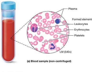
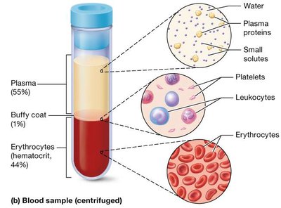
When blood is centrifuged, it separates into three layers:
Top layer: Plasma (about 55% of blood volume)
Middle layer: Buffy coat (leukocytes and platelets, about 1%)
Bottom layer: Erythrocytes (about 44%, called the hematocrit)
Plasma: Composition and Function
Plasma is a pale yellow liquid that makes up about 55% of blood volume. It is primarily water but contains proteins, nutrients, ions, gases, and waste products.
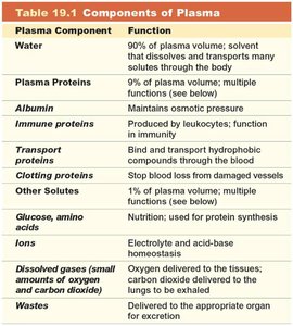
Plasma Component | Function |
|---|---|
Water | 90% of plasma volume; solvent for solutes |
Plasma Proteins | 9% of plasma volume; includes albumin, immune proteins, transport proteins, and clotting proteins |
Albumin | Maintains osmotic pressure |
Immune proteins | Produced by leukocytes; function in immunity |
Transport proteins | Bind and transport hydrophobic compounds |
Clotting proteins | Stop blood loss from damaged vessels |
Other solutes | Include nutrients, ions, dissolved gases, and wastes |
Erythrocytes (Red Blood Cells)
Structure and Function
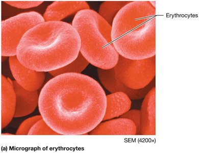
Erythrocytes are biconcave discs lacking a nucleus and most organelles, maximizing their surface area for gas exchange. Their primary function is to transport oxygen and carbon dioxide.
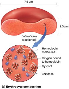
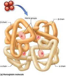
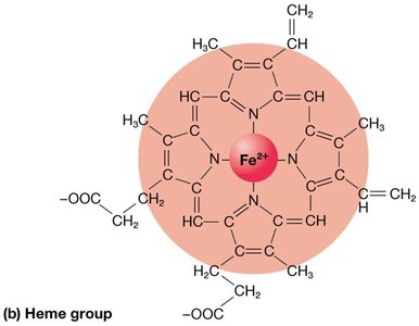
Hemoglobin: A large protein composed of four polypeptide subunits (two alpha and two beta chains), each bound to an iron-containing heme group.
Oxyhemoglobin (HbO2): Hemoglobin bound to oxygen.
Carbaminohemoglobin: Hemoglobin bound to carbon dioxide.
Carboxyhemoglobin: Hemoglobin bound to carbon monoxide (dangerous, as it prevents oxygen binding).

Erythrocytes have a lifespan of 100–120 days due to the lack of organelles for repair.
Hematopoiesis and Erythropoiesis
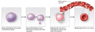
Hematopoiesis is the process of blood cell formation, occurring in red bone marrow. Erythropoiesis is the specific process that produces erythrocytes from hematopoietic stem cells (HSCs).
Hematopoietic Stem Cells (HSCs): Differentiate into all blood cell types.
Erythropoiesis: Involves several stages, including erythroblast and reticulocyte stages, before becoming mature erythrocytes.
Anemia: Causes and Types
Anemia is a condition characterized by decreased oxygen-carrying capacity of the blood. It can result from decreased hemoglobin, decreased hematocrit, or abnormal hemoglobin.
Iron Deficiency Anemia: Caused by insufficient iron for hemoglobin synthesis.
Blood Loss: Reduces hematocrit.
Pernicious Anemia: Due to vitamin B12 deficiency.
Hemolytic Anemia: Premature destruction of erythrocytes.
Aplastic Anemia: Failure of bone marrow to produce erythrocytes.
Sickle-Cell Disease: Abnormal hemoglobin causes erythrocytes to assume a sickle shape.
Leukocytes (White Blood Cells)
Types and Functions
Leukocytes are larger than erythrocytes, contain a prominent nucleus, and are involved in immune defense. They are classified as granulocytes or agranulocytes based on the presence of cytoplasmic granules.
Granulocytes: Neutrophils, eosinophils, basophils
Agranulocytes: Lymphocytes, monocytes
Neutrophils: Phagocytize bacteria and debris; most abundant (60–70%).
Eosinophils: Combat parasitic infections and modulate allergic responses (2–4%).
Basophils: Release histamine and heparin in allergic and inflammatory responses (<1%).
Lymphocytes: B cells produce antibodies; T cells mediate cellular immunity (20–25%).
Monocytes: Become macrophages in tissues; phagocytize pathogens and present antigens (3–8%).
Leukopoiesis
Leukopoiesis is the formation of leukocytes from HSCs in the bone marrow. Two cell lines are produced:
Myeloid cell line: Produces granulocytes, monocytes, erythrocytes, and platelets.
Lymphoid cell line: Produces lymphocytes (B and T cells).
Platelets and Hemostasis
Platelet Structure and Formation
Platelets are small cell fragments derived from megakaryocytes. They play a crucial role in hemostasis (blood clotting).
Thrombopoiesis: Formation of platelets from megakaryoblasts in the myeloid cell line.
Contents: Platelets contain granules with clotting factors, enzymes, mitochondria, and glycogen.
Hemostasis: The Clotting Process
Hemostasis is the process that stops blood loss from damaged vessels. It involves five steps:
Vascular Spasm: Vasoconstriction reduces blood flow.
Platelet Plug Formation: Platelets adhere to exposed collagen and aggregate.
Coagulation: Fibrin mesh forms via intrinsic and extrinsic pathways.
Clot Retraction: The clot contracts to reduce its size.
Thrombolysis: The clot is dissolved after vessel repair.
Clotting Disorders
Clotting must be tightly regulated. Disorders include:
Bleeding Disorders: Impaired clot formation leads to excessive bleeding.
Hypercoagulable Conditions: Excessive clotting increases risk of thrombosis.
Anticlot Medications: Anticoagulants, antiplatelet drugs, and thrombolytic agents are used clinically to manage clotting.
Blood Typing and Transfusion
Blood Groups and Antigens
Blood transfusions require compatibility between donor and recipient blood types. Erythrocyte surface antigens determine blood groups. The two most clinically important are the ABO and Rh groups.
ABO Group: Based on presence of A and/or B antigens.
Rh Group: Presence (Rh+) or absence (Rh−) of D antigen.
There are eight common blood types (A+, A−, B+, B−, AB+, AB−, O+, O−). Type O+ is most common in the US; AB− is least common.
Blood Typing and Matching
Blood typing uses antibodies (agglutinins) to detect antigens on erythrocytes. Agglutination indicates the presence of a specific antigen. Blood must be matched to avoid transfusion reactions, where recipient antibodies destroy donor erythrocytes, potentially causing kidney failure and death.
Universal Donor: Type O− (no A, B, or Rh antigens)
Universal Recipient: Type AB+ (no anti-A, anti-B, or anti-Rh antibodies)
Matching is always safest, even with universal types.
Clinical Laboratory Tests
Common blood tests include:
Complete Blood Count (CBC): Measures RBC, WBC, and platelet counts, hemoglobin concentration, hematocrit, and cell characteristics.
Blood Matching: Ensures compatibility for transfusions.
Clotting Time: Assesses hemostatic function.
Differential WBC Count: Determines proportions of different leukocyte types.
