 Back
BackBlood: Structure, Function, and Clinical Significance
Study Guide - Smart Notes
 Tailored notes based on your materials, expanded with key definitions, examples, and context.
Blood: Structure and Function
Overview of Blood Functions
Blood is a vital fluid connective tissue responsible for transportation, protection, and regulation within the human body. It transports oxygen, nutrients, wastes, and signaling molecules, protects against blood loss and infection, and regulates body temperature, pH, and fluid volume.
Transport: Oxygen, nutrients, wastes, hormones
Protection: Clotting (against blood loss), immune defense (against infection)
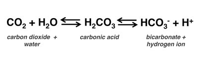
Regulation: Body temperature, pH buffering (bicarbonate system), fluid volume
Characteristics: Blood is hot (38°C), sticky, salty, metallic, and ranges from scarlet (oxygen-rich) to dark red (oxygen-poor). Its pH is slightly alkaline (7.35–7.45).
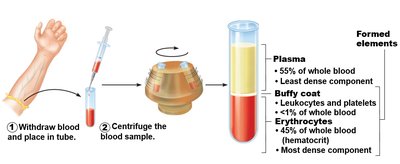
Blood Composition and Hematocrit
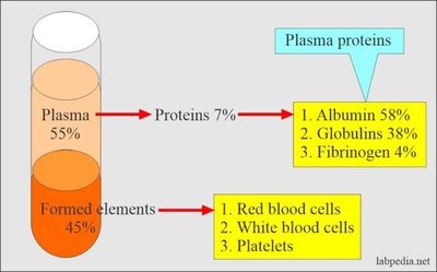
Blood is the body's only liquid connective tissue, consisting of plasma and formed elements. Hematocrit is the percentage of blood volume occupied by red blood cells (RBCs), typically 47% ± 5% for males and 42% ± 5% for females.
Plasma: Non-living fluid matrix (~90% water)
Formed Elements: Living blood cells (erythrocytes, leukocytes, platelets)
Plasma and Plasma Proteins
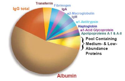
Plasma contains water, nutrients, gases, hormones, wastes, ions, and proteins. Plasma proteins (7–8% of plasma) are mostly synthesized in the liver and include:
Albumins: Most abundant, contribute to viscosity and osmolarity, influence blood pressure and fluid balance
Globulins: Provide immune functions and transport (alpha, beta, gamma types)
Fibrinogen: Precursor to fibrin, essential for blood clotting
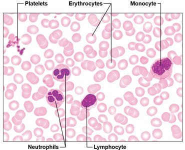
Formed Elements of Blood
Types of Formed Elements
The formed elements include erythrocytes (RBCs), leukocytes (WBCs), and platelets. Only WBCs are complete cells; RBCs lack nuclei and organelles, and platelets are cell fragments. Most formed elements originate in bone marrow and have limited lifespans.
Erythrocytes: 120 days
Platelets: 8–10 days
Leukocytes: Variable lifespan
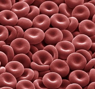
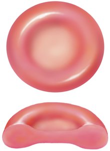
Erythrocytes (Red Blood Cells)
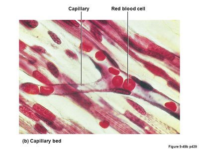
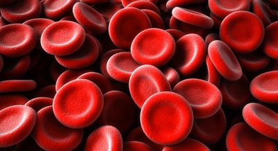
Erythrocytes are small, biconcave, non-nucleated cells specialized for oxygen transport. They do not consume the oxygen they carry, generating ATP anaerobically. Their shape increases surface area for gas exchange.
Function: Carry oxygen from lungs to tissues, transport some CO2 back to lungs
Structure: Biconcave disc, 7.5 µm diameter
Hemoglobin: 97% of cell volume (excluding water), binds reversibly with oxygen

Hemoglobin Structure and Function
Hemoglobin is a protein composed of four globin chains (2 alpha, 2 beta), each with a heme group containing iron. Each iron atom binds one oxygen molecule, allowing each hemoglobin to carry four oxygen molecules.
Color: Oxygenated blood is bright red; deoxygenated is dark red
CO2 Transport: Some CO2 binds to hemoglobin, most is transported as bicarbonate
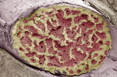
Blood Cell Production and Regulation
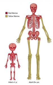
Hematopoiesis
Hematopoiesis is the production of blood cells, occurring primarily in the bone marrow. Adult humans produce billions of blood cells daily. Hematopoietic stem cells differentiate into colony-forming units (CFUs) committed to specific cell lineages.
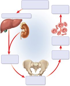
Erythropoiesis (RBC Production)
Erythropoiesis is the process of RBC formation, regulated by erythropoietin (EPO) from the kidneys in response to hypoxia. The process involves precursor cells developing ribosomes, losing organelles, and entering the bloodstream as reticulocytes before maturing into erythrocytes.
Negative Feedback: Low RBC count triggers EPO release, stimulating RBC production
Stimuli: Hypoxemia, high altitude, exercise, loss of lung tissue
Dietary Requirements for Erythropoiesis
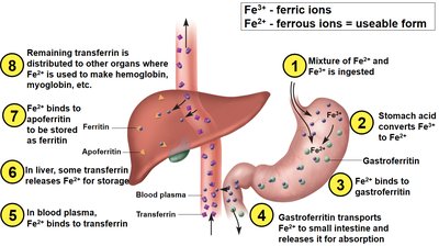
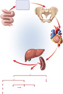
Essential nutrients for RBC production include amino acids, lipids, carbohydrates, iron, and B-complex vitamins (B12 and folic acid). Iron is stored and transported in protein complexes (ferritin, transferrin) to prevent toxicity.
Erythrocyte Death and Disposal
RBCs circulate for about 120 days before being broken down in the liver and spleen. Hemoglobin is degraded, globin is reused, iron is recycled, and heme is converted to bilirubin and excreted in bile.
Blood Disorders
Anemias
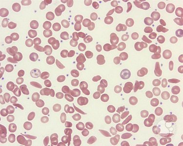
Anemia is a reduction in blood's oxygen-carrying capacity, caused by blood loss, inadequate erythropoiesis, or hemolysis. Common types include hemorrhagic, iron-deficiency, pernicious, and hemolytic anemia. Symptoms include tissue hypoxia, edema, and reduced blood viscosity.
Blood Types and Transfusion Compatibility
ABO and Rh Blood Types
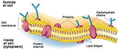
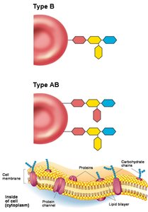
Blood types are determined by antigens (agglutinogens) on RBC membranes and antibodies (agglutinins) in plasma. The ABO system includes types A, B, AB, and O, while the Rh system is based on the presence of the D antigen.
Type A: A antigens, anti-B antibodies
Type B: B antigens, anti-A antibodies
Type AB: A and B antigens, no antibodies (universal recipient)
Type O: No antigens, both anti-A and anti-B antibodies (universal donor)
Rh+: D antigen present
Rh-: D antigen absent
Transfusion Reactions and Agglutination
Transfusion reactions occur when recipient antibodies recognize donor RBC antigens, causing agglutination (clumping) and rejection. Agglutinins in plasma bind to agglutinogens on RBCs, leading to immune responses.
Blood Type Classification Table
Blood Type | Antigens on RBC | Antibodies in Plasma | Transfusion Compatibility |
|---|---|---|---|
A | A | Anti-B | Can receive A, O |
B | B | Anti-A | Can receive B, O |
AB | A, B | None | Can receive A, B, AB, O (universal recipient) |
O | None | Anti-A, Anti-B | Can receive O (universal donor) |
Rh Group and Maternal-Fetal Incompatibility
Rh incompatibility can cause hemolytic disease of the newborn if an Rh- mother is exposed to Rh+ fetal blood. Anti-D antibodies form only after exposure, and Rhogam is administered to prevent antibody formation.
Leukocytes (White Blood Cells)
Leukocyte Functions and Types
Leukocytes are complete cells that combat disease and move from the bloodstream into tissues. They are classified as granulocytes (neutrophils, eosinophils, basophils) and agranulocytes (lymphocytes, monocytes).
Neutrophils: Phagocytize bacteria, most numerous
Eosinophils: Combat parasites, phagocytize antigen-antibody complexes
Basophils: Release histamine and heparin
Lymphocytes: T cells (destroy infected/cancerous cells), B cells (produce antibodies)
Monocytes: Differentiate into macrophages, phagocytize pathogens and debris
Leukocyte Life Cycle and Disorders
Leukopoiesis is the formation of WBCs from hematopoietic stem cells. Granulocytes and monocytes have short lifespans, while lymphocytes provide long-term immunity. Disorders include leukemia (uncontrolled proliferation) and severe combined immunodeficiency disease (SCID).
Platelets and Hemostasis
Platelet Formation and Function
Platelets are cytoplasmic fragments of megakaryocytes, essential for hemostasis. They secrete vasoconstrictors, form plugs, release clotting factors, and stimulate vessel repair.
Hemostasis: Blood Clotting Mechanism
Hemostasis involves three steps: vascular spasm (vasoconstriction), platelet plug formation, and coagulation (fibrin mesh formation). Clot retraction stabilizes the clot, and growth factors stimulate vessel repair.
Disorders of Hemostasis
Thromboembolic disorders involve undesirable clot formation (thrombus, embolus, embolism), while bleeding disorders (hemophilia) result from deficiencies in clotting factors. Hemophilia A, B, and C are treated with plasma transfusions and factor injections.
Additional info: Academic context was added to clarify mechanisms, clinical significance, and regulatory pathways.
