 Back
Blood: Structure, Function, and Disorders
Study Guide - Smart Notes
 Tailored notes based on your materials, expanded with key definitions, examples, and context.
Blood Composition and Physical Properties
Main Components of Blood
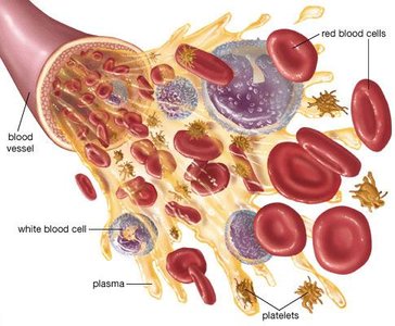
Blood is a specialized fluid connective tissue essential for transporting substances throughout the body. It consists of two main components: plasma and formed elements (erythrocytes, leukocytes, and platelets).
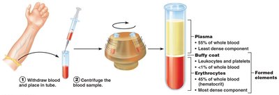
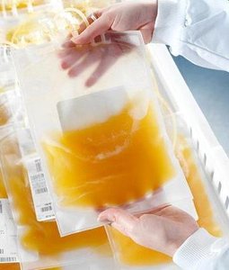
Plasma: The liquid matrix, making up about 55% of blood volume, composed mainly of water, proteins, nutrients, hormones, and waste products.
Formed Elements: Cellular components including:
Erythrocytes (Red Blood Cells, RBCs): Transport oxygen.
Leukocytes (White Blood Cells, WBCs): Immune defense.
Platelets: Cell fragments involved in blood clotting (coagulation).
Hematocrit and Blood Volume
Hematocrit is the percentage of blood volume occupied by RBCs. Normal values are approximately 47% ± 5% for males and 42% ± 5% for females. The remainder consists of plasma and a small buffy coat of leukocytes and platelets.
Blood is a sticky, opaque fluid with a pH of 7.35–7.45 and a temperature of about 38°C. It constitutes roughly 8% of body weight, with an average volume of 5–6 L in males and 4–5 L in females.
Functions of Blood
Distribution
Blood distributes:
Oxygen and nutrients to body cells
Metabolic wastes to the lungs and kidneys for elimination
Hormones from endocrine organs to target tissues
Regulation
Blood helps regulate:
Body temperature by absorbing and distributing heat
Normal pH using buffers
Adequate fluid volume in the circulatory system (osmotic pressure)
Protection
Blood protects against:
Blood loss (clotting mechanisms)
Infection (antibodies, complement proteins, and WBCs)
Blood Plasma
Composition of Plasma
Plasma is about 90% water and contains over 100 dissolved solutes, including:
Plasma proteins (mainly produced by the liver):
Albumin (60%) – maintains osmotic pressure
Globulins (36%) – transport and immune functions
Fibrinogen (4%) – blood clotting
Nitrogenous by-products (lactic acid, urea, creatinine)
Nutrients (glucose, amino acids, lipids)
Electrolytes (Na+, K+, Ca2+, Cl–, HCO3–)
Respiratory gases (O2, CO2)
Hormones
Formed Elements
Overview
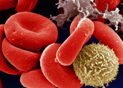
Formed elements include erythrocytes, leukocytes, and platelets. Only WBCs are complete cells; RBCs lack nuclei and organelles, and platelets are cell fragments. Most formed elements are produced in the bone marrow and have a limited lifespan.
Erythrocytes (Red Blood Cells)
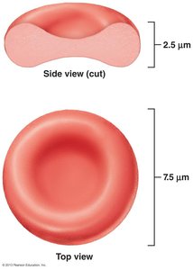
Erythrocytes are biconcave, anucleate cells specialized for oxygen transport. Their shape increases surface area for gas exchange and allows flexibility in capillaries. They are filled with hemoglobin and lack mitochondria, relying on anaerobic metabolism.
Hemoglobin Structure and Function
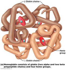
Hemoglobin (Hb) is the protein responsible for oxygen transport. Each molecule consists of four polypeptide chains (two alpha, two beta) and four heme groups, each containing an iron atom that binds one O2 molecule. Each RBC contains about 250 million Hb molecules.
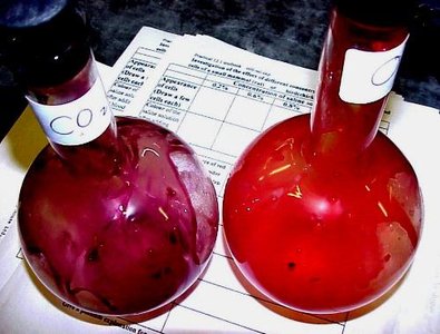
O2 loading in the lungs forms oxyhemoglobin (bright red).
O2 unloading in tissues forms deoxyhemoglobin (dark red).
CO2 loading in tissues forms carbaminohemoglobin (carries ~20% of CO2 in blood).
Hematopoiesis and Erythropoiesis
Hematopoiesis
Hematopoiesis is the process of blood cell formation, occurring primarily in red bone marrow. All formed elements arise from hematopoietic stem cells (hemocytoblasts).
Erythropoiesis
Erythropoiesis is the production of RBCs. The process involves:
Transformation of a hemocytoblast into a proerythroblast
Development through erythroblast stages (ribosome synthesis, hemoglobin accumulation, ejection of nucleus)
Formation of reticulocytes, which mature into erythrocytes
Regulation of Erythropoiesis
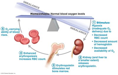
Regulated by erythropoietin (EPO), a hormone released by the kidneys in response to hypoxia (low oxygen). Adequate supplies of iron, vitamin B12, folic acid, and other nutrients are essential for effective erythropoiesis.
Fate and Destruction of Erythrocytes
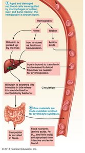
RBCs have a lifespan of 100–120 days. Old RBCs are engulfed by macrophages in the spleen, liver, and bone marrow. Hemoglobin is broken down: iron is salvaged, heme is converted to bilirubin (excreted in bile), and globin is metabolized into amino acids.
Erythrocyte Disorders
Anemia
Anemia is a condition where blood has an abnormally low oxygen-carrying capacity. It can result from insufficient erythrocytes, low hemoglobin content, or abnormal hemoglobin.
Hemorrhagic anemia: Blood loss
Hemolytic anemia: Premature RBC destruction
Aplastic anemia: Inhibition of red bone marrow
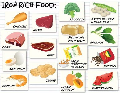
Iron-deficiency anemia: Inadequate iron intake or absorption
Pernicious anemia: Vitamin B12 deficiency
Thalassemias: Faulty globin chain synthesis
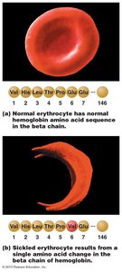
Sickle-cell anemia: Abnormal hemoglobin (HbS) causes RBCs to sickle under low oxygen

Polycythemia
Polycythemia is an excess of RBCs, increasing blood viscosity. Causes include bone marrow cancer (polycythemia vera), high altitude, or increased EPO production.
Leukocytes (White Blood Cells)
Types and Functions
Leukocytes are crucial for immune defense and are classified as granulocytes (neutrophils, eosinophils, basophils) or agranulocytes (lymphocytes, monocytes).
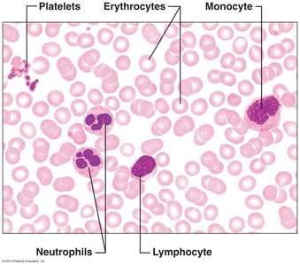
Neutrophils: Most numerous, phagocytize bacteria
Eosinophils: Digest parasitic worms, modulate immune response
Basophils: Release histamine, mediate inflammation
Lymphocytes: T cells (attack infected/tumor cells), B cells (produce antibodies)
Monocytes: Differentiate into macrophages, phagocytize pathogens and debris
Platelets and Hemostasis
Platelet Structure and Function
Platelets are fragments of megakaryocytes, essential for blood clotting. They form a temporary plug at sites of vessel injury and release chemicals that promote coagulation.
Hemostasis: Stoppage of Bleeding
Hemostasis involves three steps:
Vascular spasm (vasoconstriction)
Platelet plug formation (platelets adhere to exposed collagen and aggregate)
Coagulation (blood clotting cascade)
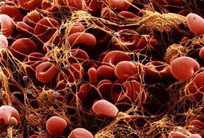
Coagulation Cascade
Coagulation transforms blood from a liquid to a gel via a cascade of clotting factors. The process includes:
Formation of prothrombin activator (intrinsic and extrinsic pathways)
Conversion of prothrombin to thrombin
Thrombin catalyzes the conversion of fibrinogen to fibrin, forming a mesh that stabilizes the clot
Blood Groups and Transfusions
ABO and Rh Blood Groups
Blood groups are determined by the presence or absence of specific antigens (agglutinogens) on RBC membranes. The most clinically significant are the ABO and Rh groups.
ABO: Types A, B, AB, O based on A and B antigens
Rh: Presence (+) or absence (–) of D antigen
Transfusion reactions can occur if mismatched blood is given, leading to agglutination and hemolysis of donor RBCs.
Selected Tables
Component | Percentage of Blood | Main Function |
|---|---|---|
Plasma | ~55% | Transport of nutrients, wastes, hormones; osmotic balance |
Erythrocytes | ~45% | Oxygen and carbon dioxide transport |
Leukocytes & Platelets (Buffy coat) | <1% | Immune defense and clotting |
Blood Disorder | Cause | Main Effect |
|---|---|---|
Anemia | Low RBCs, low Hb, abnormal Hb | Fatigue, pallor, shortness of breath |
Polycythemia | Excess RBCs | Increased blood viscosity |
Leukemia | Cancer of WBCs | Impaired immunity, anemia, bleeding |
Thrombocytopenia | Low platelets | Bleeding tendency |
Additional info: This guide expands on the original notes with definitions, examples, and tables for clarity and exam preparation.
