 Back
BackBlood: Structure, Function, and Disorders
Study Guide - Smart Notes
 Tailored notes based on your materials, expanded with key definitions, examples, and context.
Blood: Structure, Function, and Disorders
Overview of Blood Composition and Volume
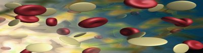
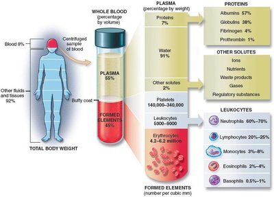
Blood is a specialized connective tissue essential for transporting substances, regulating physiological processes, and protecting the body. It consists of a liquid matrix called plasma and cellular components known as formed elements.
Plasma: The extracellular, liquid portion of blood, making up about 55% of total blood volume.
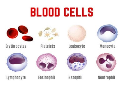
Formed Elements: Suspended cells and cell fragments, including erythrocytes (RBCs), leukocytes (WBCs), and platelets (thrombocytes).
Normal Blood Volume: 4–6 liters in adults, accounting for 7–9% of total body weight.
Blood pH and Donation
Blood is slightly alkaline, with a pH range of 7.35–7.45. Deviations toward neutrality (acidosis) can be harmful. Blood donations are critical for medical care, but stored blood has a limited shelf life (about 6 weeks).
Blood Plasma
Plasma is the liquid fraction of blood after removal of formed elements. It is primarily water (91%) with dissolved proteins, nutrients, gases, and waste products.
Major Plasma Proteins:
Albumins: Maintain osmotic pressure and water balance.
Globulins: Include antibodies for immune defense.
Fibrinogen & Prothrombin: Essential for blood clotting.
Serum: Plasma without clotting factors; contains antibodies.
Formed Elements of Blood
The formed elements include three main types: erythrocytes (RBCs), leukocytes (WBCs), and platelets (thrombocytes). Each type has distinct functions and subtypes.
RBCs (Erythrocytes): 4.2–6.2 million/mm3
WBCs (Leukocytes): 5,000–10,000/mm3
Platelets (Thrombocytes): 150,000–400,000/mm3
Hematopoiesis: Formation of Blood Cells
Hematopoiesis is the process of blood cell formation, occurring primarily in red bone marrow (myeloid tissue) and lymphoid tissues (lymph nodes, thymus, spleen). Myeloid tissue produces all blood cells except some lymphocytes and monocytes, which are formed in lymphoid tissue.
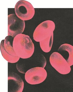
Red Blood Cells (Erythrocytes)
RBCs are specialized for oxygen and carbon dioxide transport. Their biconcave shape increases surface area for gas exchange, and the absence of a nucleus allows more space for hemoglobin (Hb).
Life Span: ~120 days
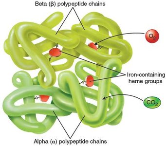
Hemoglobin: Iron-containing protein that binds O2 and CO2
Functions of RBCs
Oxygen Transport: O2 binds to hemoglobin forming oxyhemoglobin.
Carbon Dioxide Transport: CO2 binds to hemoglobin (carbaminohemoglobin) or is converted to bicarbonate.
Acid-Base Balance: RBCs help maintain pH homeostasis.
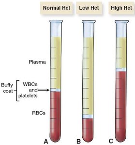
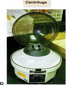
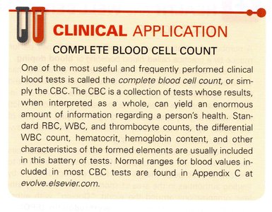
Complete Blood Count (CBC) and Hematocrit
The CBC is a comprehensive test measuring RBCs, WBCs, platelets, hemoglobin, and hematocrit (percentage of blood volume occupied by RBCs). Hematocrit is used to assess anemia or polycythemia.
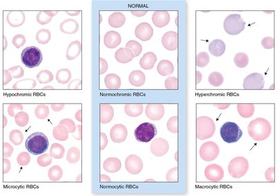
RBC Abnormalities
RBCs are classified by size and hemoglobin content:
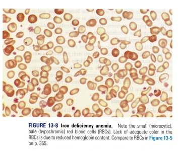
Size: Normocytes (normal), microcytic (small), macrocytic (large)
Hemoglobin Content: Normochromic (normal), hypochromic (low), hyperchromic (high)
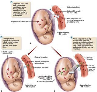
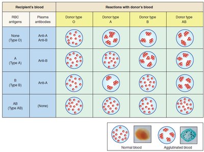
Blood Types and Transfusion Compatibility
Blood types are determined by antigens on RBCs and antibodies in plasma. The ABO and Rh systems are most clinically significant.
Type A: A antigens, anti-B antibodies
Type B: B antigens, anti-A antibodies
Type AB: A and B antigens, no antibodies (universal recipient)
Type O: No antigens, anti-A and anti-B antibodies (universal donor)
Rh System: Rh+ (antigen present), Rh– (antigen absent)
Red Blood Cell Disorders
Polycythemia
Polycythemia is an abnormal increase in RBCs, often due to bone marrow cancer. It leads to increased blood viscosity, slow flow, and risk of clotting or hemorrhage.
Anemia
Anemia is characterized by low RBC count or hemoglobin, resulting in reduced oxygen-carrying capacity. Symptoms include fatigue, pallor, and increased heart/respiratory rates.
Hemorrhagic Anemia: Due to blood loss (acute or chronic)
Aplastic Anemia: Bone marrow failure
Pernicious Anemia: Vitamin B12 deficiency
Folate Deficiency Anemia: Low folate (B9)
Iron Deficiency Anemia: Low iron, microcytic and hypochromic RBCs
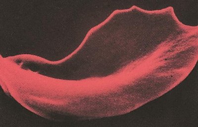
Hemolytic Anemias: Increased RBC destruction (e.g., sickle cell, thalassemia)
Leukocytes (White Blood Cells)
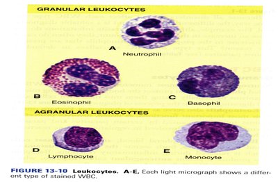
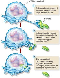
WBCs are divided into granulocytes (neutrophils, eosinophils, basophils) and agranulocytes (lymphocytes, monocytes). They play key roles in immune defense, inflammation, and phagocytosis.
Leukopenia: Low WBC count (<5,000/mm3)
Leukocytosis: High WBC count (>10,000/mm3)
White Blood Cell Disorders
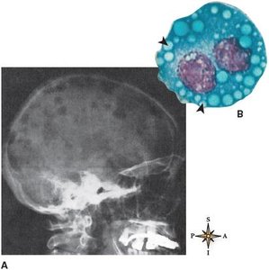
Leukemia: Cancer of WBCs, classified as acute/chronic and lymphoid/myeloid.
Multiple Myeloma: Cancer of plasma cells (B lymphocytes), causing bone lesions and anemia.
Infectious Mononucleosis: Viral infection (Epstein-Barr virus) causing atypical lymphocytosis.
Platelets and Blood Clotting
Platelets (thrombocytes) are cell fragments essential for hemostasis. They form a platelet plug at injury sites and release factors that initiate the clotting cascade.
Clotting Mechanism:
Damaged tissue releases clotting factors.
Prothrombin activator (with Ca2+) converts prothrombin to thrombin.
Thrombin converts fibrinogen to fibrin, forming a mesh that traps cells and forms a clot.
Clotting Disorders: Hemophilia (factor VIII deficiency), thrombocytopenia (low platelets), vitamin K deficiency.
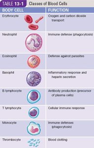
Summary Table: Classes of Blood Cells and Functions
Body Cell | Function |
|---|---|
Erythrocyte | Oxygen and carbon dioxide transport |
Neutrophil | Immune defense (phagocytosis) |
Eosinophil | Defense against parasites |
Basophil | Inflammatory response and heparin secretion |
B lymphocyte | Antibody production; precursor of plasma cells |
T lymphocyte | Cellular immune response |
Monocyte | Immune defense (phagocytosis) |
Thrombocyte | Blood clotting |
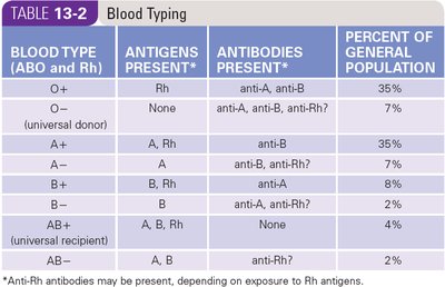
Summary Table: Blood Typing
Blood Type (ABO and Rh) | Antigens Present | Antibodies Present | Percent of General Population |
|---|---|---|---|
O+ | Rh | anti-A, anti-B | 35% |
O– | None | anti-A, anti-B, anti-Rh? | 7% |
A+ | A, Rh | anti-B | 35% |
A– | A | anti-B, anti-Rh? | 7% |
B+ | B, Rh | anti-A | 8% |
B– | B | anti-A, anti-Rh? | 2% |
AB+ | A, B, Rh | None | 4% |
AB– | A, B | anti-Rh? | 2% |
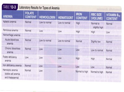
Summary Table: Laboratory Results for Types of Anemia
Anemia | Folate Content | Hemoglobin | Hematocrit | Iron Content | RBC Size (Volume) | Vitamin B12 Content |
|---|---|---|---|---|---|---|
Aplastic anemia | Normal | Low to normal | Low to normal | Normal to high | Normal to slightly high | Normal |
Pernicious anemia | Normal | Low | Low | Normal | High | Low |
Hemorrhagic anemia | Normal | Low | Low | High | Normal | Normal |
Acute blood-loss anemia | Normal | Low | Low | Normal | Slightly low | Normal |
Chronic blood-loss anemia | Normal | Low | Low | Low | Low to normal | Normal |
Folate deficiency anemia | Low | Low | Low | Normal | Low | Normal |
Iron deficiency anemia | Normal | Low | Low | Low | Low | Normal |
Hemolytic anemia (sickle cell anemia and thalassemia) | Normal | Low | Low | Normal to high | Low | Normal |
Key Equations
Hematocrit (Hct):
Oxygen Carrying Capacity:
Additional info:
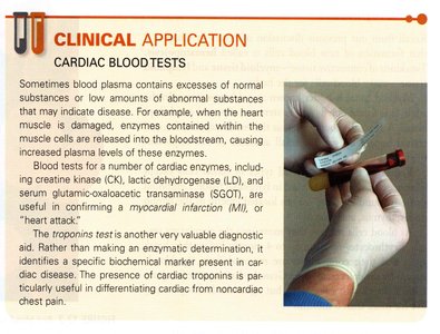
Blood disorders can be diagnosed using laboratory tests such as CBC, blood smears, and specific biochemical markers.
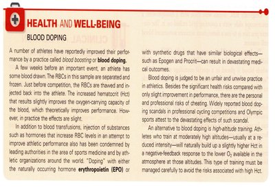
Blood doping and erythropoietin (EPO) use can artificially increase RBC count but pose health risks.
