 Back
BackBlood: Structure, Function, and Disorders
Study Guide - Smart Notes
 Tailored notes based on your materials, expanded with key definitions, examples, and context.
Blood: Structure, Function, and Disorders
Introduction to Blood
Blood is a specialized connective tissue essential for transporting substances, regulating physiological processes, and protecting the body against disease. It consists of a liquid matrix called plasma and various formed elements, including red blood cells (RBCs), white blood cells (WBCs), and platelets.
Blood Composition and Volume
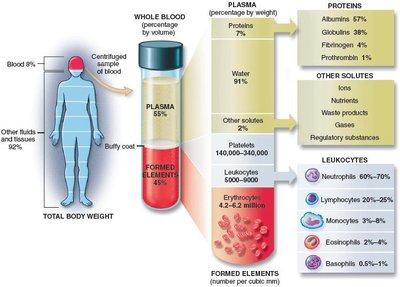
Plasma: The liquid fraction of blood, making up about 55% of total blood volume. It contains water, proteins, nutrients, gases, and waste products.
Formed Elements: Suspended in plasma, these include RBCs, WBCs, and platelets, comprising about 45% of blood volume.
Normal Blood Volumes: Whole blood volume averages 4–6 L in adults, accounting for 7–9% of total body weight.
Blood pH and Donation
Blood pH: Normally ranges from 7.35 to 7.45, making blood slightly alkaline. A decrease toward neutral is termed acidosis.
Blood Donation: Approximately 14 million units are donated annually. Plasma expanders can temporarily maintain blood volume after hemorrhage, but donated blood can only be stored for up to 6 weeks.
Blood Plasma
Plasma is the liquid portion of blood after removal of formed elements. It is composed mainly of water (91%) and dissolved substances such as proteins, nutrients, gases, and waste products.
Plasma Proteins:
Albumins: Maintain osmotic pressure and water balance.
Globulins: Include antibodies for immune defense.
Fibrinogen and Prothrombin: Essential for blood clotting.
Serum: Plasma without clotting factors; contains antibodies.
Formed Elements of Blood
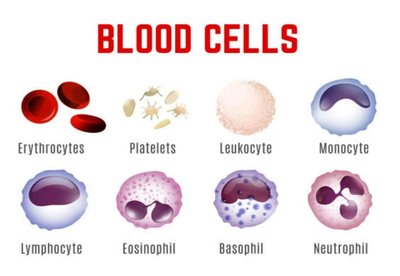
The formed elements include three main types:
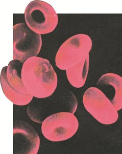
Red Blood Cells (Erythrocytes)
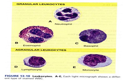
White Blood Cells (Leukocytes)
Granular Leukocytes: Neutrophils, eosinophils, basophils
Agranular Leukocytes: Lymphocytes, monocytes
Platelets (Thrombocytes)
Numbers of Formed Elements
RBCs: 4.2–6.2 million/mm3
WBCs: 5,000–10,000/mm3
Platelets: 150,000–400,000/mm3
Hematopoiesis
Hematopoiesis is the process of blood cell formation, occurring primarily in red bone marrow (myeloid tissue) and lymphoid tissue (lymph nodes, thymus, spleen). Most blood cells are produced in red bone marrow, except some lymphocytes and monocytes, which are formed in lymphoid tissue.
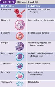
Classes of Blood Cells and Their Functions
Body Cell | Function |
|---|---|
Erythrocyte | Oxygen and carbon dioxide transport |
Neutrophil | Immune defense (phagocytosis) |
Eosinophil | Defense against parasites |
Basophil | Inflammatory response and heparin secretion |
B lymphocyte | Antibody production; precursor of plasma cells |
T lymphocyte | Cellular immune response |
Monocyte | Immune defense (phagocytosis) |
Thrombocyte | Blood clotting |
Red Blood Cells (Erythrocytes)
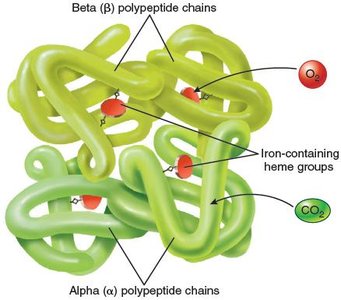
RBCs are biconcave, anucleate cells specialized for gas transport. Their shape increases surface area for gas exchange and allows flexibility to pass through capillaries. The absence of a nucleus provides more space for hemoglobin (Hb), the oxygen-carrying protein.
Hemoglobin and Gas Transport
Oxyhemoglobin: Hemoglobin bound to oxygen
Carbaminohemoglobin: Hemoglobin bound to carbon dioxide
RBCs also help maintain acid-base balance by converting CO2 to bicarbonate
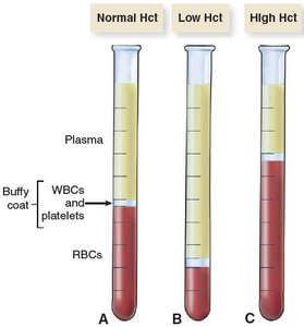
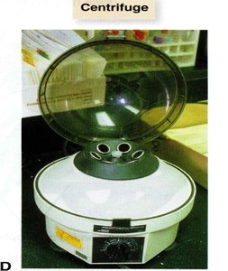
Hematocrit and Blood Testing
The hematocrit (Hct) is the percentage of blood volume occupied by RBCs. It is measured using a centrifuge, which separates blood into plasma, buffy coat (WBCs and platelets), and RBCs.
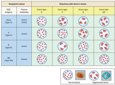
Blood Types and Transfusion Compatibility
Blood types are determined by the presence or absence of specific antigens on RBC membranes. The two main systems are ABO and Rh.
ABO System:
Type A: A antigens, anti-B antibodies
Type B: B antigens, anti-A antibodies
Type AB: A and B antigens, no antibodies (universal recipient)
Type O: No antigens, anti-A and anti-B antibodies (universal donor)
Rh System:
Rh-positive: Rh antigen present
Rh-negative: No Rh antigen; anti-Rh antibodies develop after exposure
BLOOD TYPE (ABO and Rh) | ANTIGENS PRESENT | ANTIBODIES PRESENT | PERCENT OF GENERAL POPULATION |
|---|---|---|---|
O+ | Rh | anti-A, anti-B | 35% |
O− | None | anti-A, anti-B, anti-Rh? | 7% |
A+ | A, Rh | anti-B | 35% |
A− | A | anti-B, anti-Rh? | 7% |
B+ | B, Rh | anti-A | 8% |
B− | B | anti-A, anti-Rh? | 2% |
AB+ | A, B, Rh | None | 4% |
AB− | A, B | anti-Rh? | 2% |
Red Blood Cell Disorders
Polycythemia
Excessive RBC production, often due to bone marrow cancer
Symptoms: Increased blood viscosity, slow flow, hypertension, risk of clotting
Treatment: Blood removal, chemotherapy, irradiation
Anemia
Low RBC count or abnormal hemoglobin reduces oxygen-carrying capacity
Symptoms: Fatigue, pallor, weakness, increased heart and respiratory rates
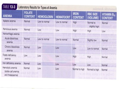
Types of Anemia
Anemia | Folate Content | Hemoglobin | Hematocrit | Iron Content | RBC Size | Vitamin B12 Content |
|---|---|---|---|---|---|---|
Aplastic | Normal | Low | Low | Normal | Low | Normal |
Pernicious | Low to normal | Low | Low | Normal | High | Low |
Hemorrhagic | Normal | Low | Low | High | Normal | Normal |
Iron deficiency | Normal | Low | Low | Low | Low | Normal |
Hemolytic | Normal | Low | Low | Normal to high | Low | Normal |
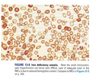
Iron Deficiency Anemia
Caused by insufficient iron for hemoglobin synthesis
RBCs are microcytic and hypochromic
Treatment: Oral iron supplements
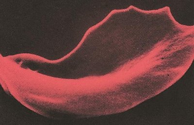
Sickle Cell Anemia
Genetic disorder causing abnormal hemoglobin (HbS)
RBCs become sickle-shaped under low oxygen, leading to hemolysis and vascular blockage
White Blood Cells (Leukocytes)
WBCs are divided into granulocytes (neutrophils, eosinophils, basophils) and agranulocytes (lymphocytes, monocytes). They play key roles in immune defense, inflammation, and phagocytosis.
Leukocyte Disorders
Leukopenia: Low WBC count, often due to bone marrow failure or immune diseases
Leukocytosis: High WBC count, common in infections and leukemia
Leukemia: Cancer of WBCs, classified as acute or chronic, lymphocytic or myeloid
Platelets and Blood Clotting
Platelets (thrombocytes) are cell fragments essential for blood clotting. They form a platelet plug at injury sites and release factors that initiate the coagulation cascade.
Clotting Mechanism:
Damaged tissue releases clotting factors, forming prothrombin activator
Prothrombin activator and calcium convert prothrombin to thrombin
Thrombin converts fibrinogen to fibrin, forming a mesh that traps blood cells
Clotting Disorders
Hemophilia: X-linked disorder causing deficiency of clotting factor VIII; leads to excessive bleeding
Thrombocytopenia: Low platelet count, causing bleeding and purpura
Vitamin K Deficiency: Impairs synthesis of clotting factors
Summary Table: Blood Cell Types and Functions
Cell Type | Main Function |
|---|---|
Erythrocyte | Oxygen and carbon dioxide transport |
Neutrophil | Phagocytosis of bacteria |
Eosinophil | Defense against parasites |
Basophil | Release histamine and heparin |
Lymphocyte | Immune response (B and T cells) |
Monocyte | Phagocytosis; become macrophages |
Platelet | Blood clotting |
Key Equations
Hematocrit (Hct):
Oxygen Transport:
Carbon Dioxide Transport:
Clinical Applications
Complete Blood Cell Count (CBC): Measures RBC, WBC, platelet counts, hemoglobin, hematocrit, and more to assess health status.
Blood Typing: Essential for safe transfusions and organ transplantation.
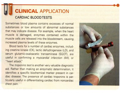
Cardiac Blood Tests: Enzyme levels (e.g., troponins) help diagnose myocardial infarction.
Review Questions
Blood pH is between 7.35 and 7.45. This makes the blood:
B. Slightly alkaline
The formed element that functions in oxygen and carbon dioxide transport is the:
A. Erythrocyte
During periods of chronic blood loss, the body helps maintain homeostasis by producing:
C. Normocytic RBCs
If you have type A blood, type ____ antigen is on the RBC and the plasma contains _____ antibodies.
C. A, anti-B
_____ anemia results from a deficiency of vitamin B12.
D. Pernicious
_____ leukemia results from cancerous transformation of granulocytic precursor cells in the bone marrow.
B. Chronic myeloid
A common type of clotting disorder resulting in a decrease in the platelet count is called:
D. Thrombocytopenia
