 Back
BackBlood: Structure, Function, and Disorders
Study Guide - Smart Notes
 Tailored notes based on your materials, expanded with key definitions, examples, and context.
Blood: Structure, Function, and Disorders
Introduction to Blood
Blood is a specialized connective tissue essential for transporting substances, regulating physiological processes, and protecting the body against disease. It consists of a liquid matrix called plasma and various formed elements, including red blood cells, white blood cells, and platelets.
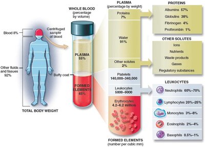
Blood Composition and Volume
Plasma: The liquid fraction of blood, making up about 55% of total blood volume. It contains water, proteins, nutrients, gases, and waste products.
Formed Elements: Suspended in plasma, these include erythrocytes (RBCs), leukocytes (WBCs), and platelets (thrombocytes).
Normal Blood Volumes: Plasma: 2.6 L; Formed elements: 2.4 L; Whole blood: 4–6 L (7–9% of body weight).
Blood pH and Donation
pH: Blood is slightly alkaline, with a pH of 7.35–7.45. A decrease toward neutral is called acidosis.
Donation: About 14 million units are donated annually. Plasma expanders can temporarily maintain volume after hemorrhage, but stored blood is viable for only 6 weeks.
Blood Plasma
Plasma is the liquid portion of blood minus the formed elements. It is composed mainly of water (91%) and dissolved substances such as nutrients, salts, gases, and proteins.
Plasma Proteins:
Albumins: Maintain osmotic pressure and water balance.
Globulins: Include antibodies for immune defense.
Fibrinogen and Prothrombin: Essential for blood clotting.
Serum: Plasma minus clotting factors; contains antibodies.
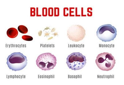
Formed Elements of Blood
Red Blood Cells (Erythrocytes): 4.2–6.2 million/mm3
White Blood Cells (Leukocytes): 5,000–10,000/mm3
Platelets (Thrombocytes): 150,000–400,000/mm3
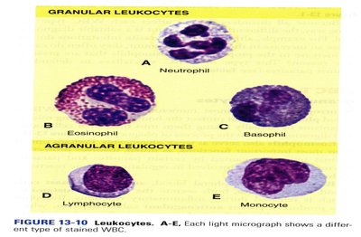
Granular Leukocytes: Neutrophils, eosinophils, basophils
Agranular Leukocytes: Lymphocytes, monocytes
Hematopoiesis
Hematopoiesis is the process of blood cell formation, occurring in myeloid (red bone marrow) and lymphoid tissues (lymph nodes, thymus, spleen). Most blood cells are formed in red bone marrow, except some lymphocytes and monocytes, which are produced in lymphoid tissue.
Mechanisms of Blood Disease
Blood diseases often result from failure of myeloid or lymphoid tissues due to toxins, radiation, genetic defects, nutritional deficiencies, or cancers (e.g., leukemia).
Aspiration biopsy cytology (ABC) is used to diagnose blood diseases by examining blood-forming tissues.
Red Blood Cells (Erythrocytes)
Structure and Function
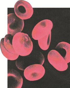
RBCs are biconcave disks with flexible membranes, lacking nuclei and most organelles, which maximizes space for hemoglobin (Hb). Their primary function is to transport oxygen and carbon dioxide.
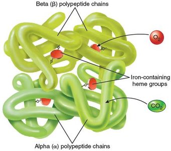
Hemoglobin (Hb): The red pigment that binds O2 (as oxyhemoglobin) and CO2 (as carbaminohemoglobin).
Lifespan: About 120 days.
RBC Count and Hematocrit
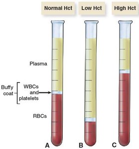
The complete blood cell count (CBC) measures various blood constituents, including RBCs. Hematocrit (PCV) is the percentage of blood volume occupied by RBCs.
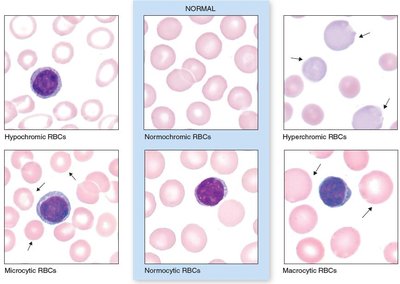
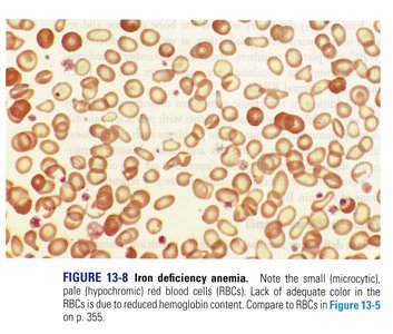
RBC Abnormalities
Size: Normocytes (normal), microcytic (small), macrocytic (large)
Hemoglobin Content: Normochromic (normal), hypochromic (low), hyperchromic (high)
Blood Types and Transfusion
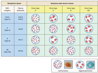
ABO System
Type A: A antigens, anti-B antibodies
Type B: B antigens, anti-A antibodies
Type AB: A and B antigens, no antibodies (universal recipient)
Type O: No antigens, anti-A and anti-B antibodies (universal donor)
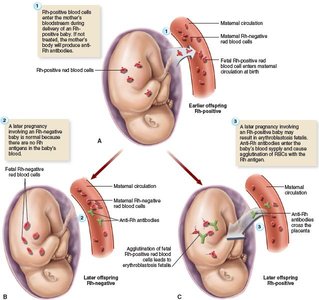
Rh System
Rh-positive: Rh antigen present
Rh-negative: No Rh antigen; anti-Rh antibodies form only after exposure
Erythroblastosis fetalis: Hemolytic disease of the newborn due to Rh incompatibility
Red Blood Cell Disorders
Polycythemia
Excessive RBC production, often due to bone marrow cancer
Symptoms: Increased blood viscosity, slow flow, hypertension, risk of clotting
Treatment: Blood removal, chemotherapy, irradiation
Anemia
Anemia is characterized by low RBC count or abnormal hemoglobin, resulting in reduced oxygen-carrying capacity. Normal Hb: 12–14 g/100 mL; anemia: <9 g/100 mL.
Symptoms: Fatigue, pallor, weakness, increased heart and respiratory rates
Types of Anemia
Hemorrhagic: Due to blood loss (acute or chronic)
Aplastic: Bone marrow failure (toxins, radiation, drugs)
Pernicious: Vitamin B12 deficiency, macrocytic RBCs, CNS symptoms
Folate Deficiency: Low folate, common in malnutrition/alcoholism
Iron Deficiency: Microcytic, hypochromic RBCs, low hematocrit
Hemolytic: Increased RBC destruction (e.g., sickle cell, thalassemia)
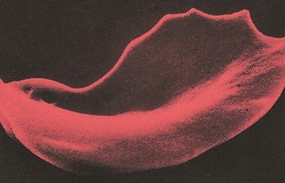
Sickle Cell Anemia
Genetic disorder causing abnormal hemoglobin (HbS)
RBCs sickle under low O2, leading to hemolysis, pain crises, and organ damage
Thalassemia
Inherited hemolytic anemia, common in Mediterranean populations
Microcytic, short-lived RBCs; severe forms require marrow/stem cell transplantation
Hemolytic Disease of the Newborn (Erythroblastosis Fetalis)
Caused by maternal-fetal blood incompatibility (ABO or Rh)
Symptoms: Jaundice, anemia, organ damage; prevention with RhoGAM
White Blood Cells (Leukocytes)
Types and Functions
Granulocytes:
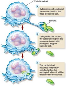
Neutrophils: Most numerous, phagocytic, increase in bacterial infections
Eosinophils: Defend against parasites, involved in allergies
Basophils: Release histamine (inflammation) and heparin (anticoagulant)
Agranulocytes:
Lymphocytes: B cells (antibody production), T cells (cellular immunity)
Monocytes: Largest WBCs, become macrophages in tissues
WBC Disorders
Leukopenia: Low WBC count (<5,000/mm3), seen in immune disorders (e.g., AIDS)
Leukocytosis: High WBC count (>10,000/mm3), common in infections and leukemia
Differential WBC Count: Measures proportions of each WBC type
Leukemias and Blood Cancers
Lymphoid Neoplasms: From B/T lymphocyte precursors
Myeloid Neoplasms: From granulocyte, monocyte, RBC, or platelet precursors
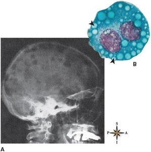
Multiple Myeloma: Cancer of plasma cells, causes bone lesions and anemia
Leukemia Types:
Chronic Lymphocytic Leukemia (CLL): Older adults, slow progression
Acute Lymphocytic Leukemia (ALL): Children, rapid onset, high cure rate in children
Chronic Myeloid Leukemia (CML): Adults, slow progression, treatable with Gleevec
Acute Myeloid Leukemia (AML): Adults, rapid progression, poor prognosis
Infectious Mononucleosis
Viral infection (Epstein-Barr virus), common in young adults
Symptoms: Fever, fatigue, sore throat, lymphadenopathy
Platelets and Blood Clotting
Platelet Function
Platelets (thrombocytes) are essential for hemostasis (stopping bleeding).
They form a platelet plug at injury sites and release clotting factors.
Clotting Mechanism
Damaged tissues release clotting factors, forming prothrombin activator.
Prothrombin activator and calcium convert prothrombin to thrombin.
Thrombin converts fibrinogen to fibrin, forming a mesh that traps blood cells to form a clot.
Clotting Disorders
Thrombus: Stationary clot
Embolus: Circulating clot
Hemophilia: X-linked disorder, lack of factor VIII, causes severe bleeding
Thrombocytopenia: Low platelet count, causes bleeding and purpura
Vitamin K Deficiency: Impairs synthesis of clotting factors
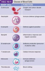
Summary Table: Classes of Blood Cells
Body Cell | Function |
|---|---|
Erythrocyte | Oxygen and carbon dioxide transport |
Neutrophil | Immune defense (phagocytosis) |
Eosinophil | Defense against parasites |
Basophil | Inflammatory response and heparin secretion |
B lymphocyte | Antibody production (precursor of plasma cells) |
T lymphocyte | Cellular immune response |
Monocyte | Immune defense (phagocytosis) |
Thrombocyte | Blood clotting |
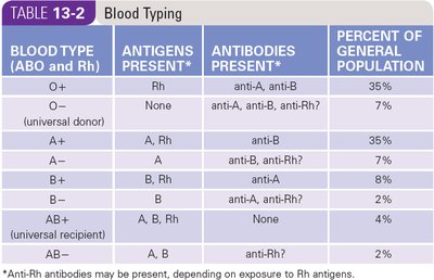
Summary Table: Blood Typing
Blood Type (ABO and Rh) | Antigens Present | Antibodies Present | Percent of Population |
|---|---|---|---|
O+ | Rh | anti-A, anti-B | 35% |
O− | None | anti-A, anti-B, anti-Rh? | 7% |
A+ | A, Rh | anti-B | 35% |
A− | A | anti-B, anti-Rh? | 7% |
B+ | B, Rh | anti-A | 8% |
B− | B | anti-A, anti-Rh? | 2% |
AB+ | A, B, Rh | None | 4% |
AB− | A, B | anti-Rh? | 2% |
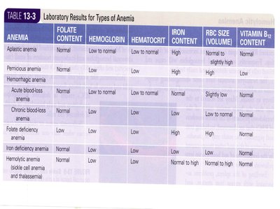
Summary Table: Laboratory Results for Types of Anemia
Anemia | Folate Content | Hemoglobin | Hematocrit | Iron Content | RBC Size (Volume) | Vitamin B12 Content |
|---|---|---|---|---|---|---|
Aplastic anemia | Normal | Low | Low | Normal to high | Normal to slightly high | Normal |
Pernicious anemia | Low to normal | Low | Low | Normal | High | Low |
Hemorrhagic anemia | Normal | Low | Low | High | Normal | Normal |
Acute blood-loss anemia | Normal | Low | Low | Normal | Slightly low | Normal |
Chronic blood-loss anemia | Normal | Low | Low | Low | Low | Normal |
Folate deficiency anemia | Low | Low | Low | Normal | High | Normal |
Iron deficiency anemia | Normal | Low | Low | Low | Low | Normal |
Hemolytic anemia (sickle cell, thalassemia) | Normal | Low | Low | Normal to high | Low | Normal |
Key Equations
Hematocrit (Hct):
Oxygen Transport:
CO2 Transport:
Clinical Applications
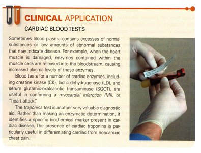
Cardiac Blood Tests: Enzyme levels (CK, LDH, SGOT) and troponins help diagnose myocardial infarction.
Complete Blood Cell Count (CBC): Provides information on RBC, WBC, platelet counts, hemoglobin, hematocrit, and more.
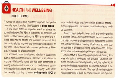
Health and Well-Being: Blood Doping
Blood doping involves increasing RBC count to enhance athletic performance, often by transfusion or using erythropoietin (EPO). It is considered unsafe and unethical due to risks of high hematocrit, blood viscosity, and cardiovascular complications.
Review Questions
Blood pH is between 7.35 and 7.45. This makes the blood: B. Slightly alkaline
The formed element that functions in oxygen and carbon dioxide transport is the: A. Erythrocyte
During periods of chronic blood loss, the body helps maintain homeostasis by producing: C. Normocytic RBCs
If you have type A blood, type ____ antigen is on the RBC and the plasma contains _____ antibodies. C. A, anti-B
_____ anemia results from a deficiency of vitamin B12. D. Pernicious
_____ leukemia results from cancerous transformation of granulocytic precursor cells in the bone marrow. B. Chronic myeloid
A common type of clotting disorder resulting in a decrease in the platelet count is called: D. Thrombocytopenia
