 Back
BackBlood: Structure, Function, and Disorders – ANP College Study Guide
Study Guide - Smart Notes
 Tailored notes based on your materials, expanded with key definitions, examples, and context.
Blood: Internal Transport System
Overview and Functions
Blood is a life-sustaining fluid that circulates within a closed cardiovascular system in vertebrates. It is essential for the transport of substances, regulation of physiological parameters, and protection against disease.
Transport: Delivers oxygen and nutrients to cells, removes metabolic wastes, and transports hormones.
Regulation: Maintains body temperature, pH balance (using buffers and bicarbonate ions), and fluid volume.
Protection: Prevents blood loss (clot formation) and infection (immune agents such as antibodies, complement proteins, and white blood cells).
Composition of Blood
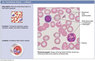
Blood as Connective Tissue
Blood is the only fluid tissue in the body, classified as a connective tissue. It consists of a nonliving matrix (plasma) and living cells (formed elements).
Plasma: The fluid matrix, making up about 55% of blood volume.
Formed Elements: Suspended in plasma; include erythrocytes (RBCs), leukocytes (WBCs), and platelets (thrombocytes).
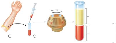
Major Components of Whole Blood
When blood is centrifuged, it separates into three layers:
Erythrocytes: Bottom layer (~45% of blood, hematocrit).
Buffy Coat: Middle layer (<1%, contains WBCs and platelets).
Plasma: Top layer (~55%).
Physical Characteristics & Volume
Blood is sticky, opaque, and has a metallic taste. Its color varies with oxygen content (scarlet red when oxygenated, dark red when deoxygenated). Blood makes up about 8% of body weight, with average volumes of 5–6 L in males and 4–5 L in females.
Distribution: Lungs (20%), veins (60%), heart/arteries/capillaries (20%).
Blood Plasma
Composition and Functions
Plasma is a straw-colored, sticky fluid composed of about 90% water and over 100 dissolved solutes, including nutrients, gases, hormones, wastes, proteins, and inorganic ions.
Plasma Proteins: Most abundant solutes, produced mainly by the liver. Albumin makes up 60% of plasma proteins and functions in coagulation, immune response, molecule transport, osmotic pressure, and buffering.
Constituent | Description and Importance |
|---|---|
Nonprotein nitrogenous substances | By-products of metabolism (urea, uric acid, creatinine, ammonium salts) |
Nutrients (organic) | Glucose, amino acids, fatty acids, glycerol, triglycerides, cholesterol, vitamins |
Respiratory gases | Oxygen and carbon dioxide |
Hormones | Steroid and thyroid hormones carried by plasma proteins |

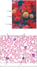
Formed Elements
Types and Characteristics
Formed elements include RBCs, WBCs, and platelets. Only WBCs are complete cells; RBCs lack nuclei and organelles, and platelets are cell fragments. Most formed elements survive only a few days and originate in bone marrow.
Erythrocytes (Red Blood Cells)
Structure and Function
Erythrocytes are small, biconcave, anucleate cells specialized for gas transport. Their shape provides a large surface area for gas exchange, and they are filled with hemoglobin (Hb).
Hemoglobin: Binds reversibly with oxygen; normal values are 13–18 g/100mL (males) and 12–16 g/100mL (females).
Structure: Composed of four polypeptide chains (two alpha, two beta) and four heme groups, each with a central iron atom that binds one oxygen molecule.
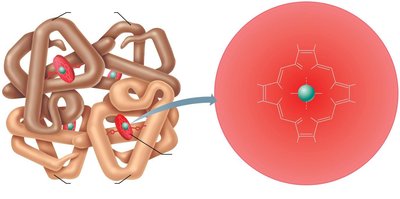
Hemoglobin Function
Each Hb molecule can transport four O2 molecules.
O2 loading in lungs produces oxyhemoglobin (ruby red).
O2 unloading in tissues produces deoxyhemoglobin (dark red).
CO2 loading in tissues produces carbaminohemoglobin (20% of CO2 in blood).
Production and Regulation of Erythrocytes
Hematopoiesis and Erythropoiesis
Hematopoiesis is the formation of all blood cells, occurring in red bone marrow. Erythropoiesis is the specific process of RBC formation, taking about 15 days and involving several stages from stem cell to mature erythrocyte.
Regulation: Controlled by erythropoietin (EPO), a hormone released by kidneys in response to hypoxia.
Dietary Requirements: Amino acids, lipids, carbohydrates, iron, vitamin B12, and folic acid are essential for erythropoiesis.
Fate and Destruction of Erythrocytes
Life Cycle and Breakdown
RBCs have a lifespan of 100–120 days. Senescent RBCs are broken down in the spleen, liver, and bone marrow. Hemoglobin is separated into heme, iron, and globin:
Iron: Stored as ferritin or hemosiderin, reused for erythropoiesis.
Heme: Degraded to bilirubin, excreted in bile, and further metabolized to stercobilin (excreted in feces).
Globin: Metabolized into amino acids.
Erythrocyte Disorders
Anemia
Anemia is a condition of abnormally low O2-carrying capacity. It is classified by cause:
Blood Loss: Hemorrhagic anemia (acute or chronic).
Not Enough RBCs Produced: Iron-deficiency anemia, pernicious anemia (B12 deficiency), renal anemia (lack of EPO), aplastic anemia (bone marrow destruction).
Too Many RBCs Destroyed: Hemolytic anemias (incompatible transfusions, infections, genetic disorders such as thalassemias and sickle-cell anemia).
Polycythemia
Polycythemia is an abnormal excess of RBCs, increasing blood viscosity and causing sluggish flow. Causes include bone marrow cancer (polycythemia vera), high altitude, and blood doping.
Leukocytes (White Blood Cells)
Structure and Function
Leukocytes are complete cells with nuclei and organelles, making up less than 1% of blood volume. They defend against disease and can leave capillaries via diapedesis.
Leukocytosis: WBC count over 11,000/μL, normal response to infection.
Categories: Granulocytes (neutrophils, eosinophils, basophils) and agranulocytes (lymphocytes, monocytes).
Types and Relative Percentages
Type | Percentage |
|---|---|
Neutrophils | 50–70% |
Lymphocytes | 25–45% |
Monocytes | 3–8% |
Eosinophils | 2–4% |
Basophils | 0.5–1% |
Granulocytes
Neutrophils: Most numerous, phagocytic, kill microbes by respiratory burst.
Eosinophils: Digest parasitic worms, modulate immune response, involved in allergies and asthma.
Basophils: Rarest, contain histamine (inflammatory mediator), functionally similar to mast cells.
Agranulocytes
Lymphocytes: Crucial to immunity; T cells (cell-mediated), B cells (antibody production).
Monocytes: Largest WBCs, differentiate into macrophages, phagocytic, activate lymphocytes.
Production and Life Span of Leukocytes
Leukopoiesis
Leukopoiesis is stimulated by interleukins and colony-stimulating factors (CSFs). All leukocytes originate from hemocytoblast stem cells, branching into lymphoid (lymphocytes) and myeloid (other elements) pathways.
Leukocyte Disorders
Leukemias and Infectious Mononucleosis
Leukemias: Cancerous overproduction of abnormal WBCs; classified as myeloid or lymphocytic, acute or chronic.
Infectious Mononucleosis: Viral disease caused by Epstein-Barr virus, results in excessive lymphocytes.
Platelets
Structure and Function
Platelets are fragments of megakaryocytes, involved in clotting. They form temporary plugs in vessel breaks and are regulated by thrombopoietin.
Normal Count: 150,000–400,000 platelets/mL.
Life Span: About 10 days.
Hemostasis
Steps in Hemostasis
Hemostasis is the process of stopping bleeding, involving three steps:
Vascular Spasm: Vasoconstriction in response to injury.
Platelet Plug Formation: Platelets adhere to exposed collagen, become activated, and release chemical messengers (ADP, serotonin, thromboxane A2).
Coagulation: Reinforces plug with fibrin threads; involves intrinsic and extrinsic pathways, leading to activation of factor X and formation of prothrombin activator.
Coagulation Pathways
Intrinsic Pathway: Triggered by factors within blood.
Extrinsic Pathway: Triggered by tissue factor outside blood.
Common Pathway: Thrombin converts fibrinogen to fibrin, forming a mesh that stabilizes the clot.
Clot Retraction and Fibrinolysis
Clot Retraction: Platelets contract, pulling fibrin strands and drawing vessel edges together.
Fibrinolysis: Plasminogen is converted to plasmin, which digests fibrin and dissolves the clot.
Disorders of Hemostasis
Thromboembolic and Bleeding Disorders
Thromboembolic Disorders: Undesirable clot formation (thrombus, embolus).
Bleeding Disorders: Impaired clot formation (hemophilia, liver dysfunction, vitamin K deficiency).
DIC: Disseminated intravascular coagulation involves both clotting and bleeding.
Blood Transfusions
Transfusion Principles
Blood transfusions are used to restore blood volume and oxygen-carrying capacity. Blood typing is essential to prevent fatal transfusion reactions.
ABO and Rh Groups: Major antigens on RBCs; mismatched transfusions cause agglutination and hemolysis.
Universal Donor: Type O- (no A, B, or Rh antigens).
Universal Recipient: Type AB+ (no anti-A, anti-B, or anti-Rh antibodies).
Diagnostic Blood Tests
Clinical Applications
Hematocrit: Indicates anemia.
Blood Glucose: Checks for diabetes.
Differential WBC Count: Helps diagnose infections.
CMP and CBC: Assess overall health and detect disorders.
Developmental Aspects of Blood
Fetal and Aging Blood
Fetal Blood: Forms in yolk sac, liver, and spleen; hemoglobin F has higher O2 affinity.
Aging: Blood diseases often result from disorders of heart, vessels, or immune system.
