 Back
Blood: Structure, Function, and Hemostasis
Study Guide - Smart Notes
 Tailored notes based on your materials, expanded with key definitions, examples, and context.
Blood: Structure and Function
Functions of Blood
Blood is a vital connective tissue that serves as the internal transport system of the body. Its functions are categorized into transport, regulation, and protection.
Transport: Blood delivers oxygen and nutrients to body cells, removes metabolic wastes for elimination, and transports hormones from endocrine organs to target tissues.
Regulation: Blood maintains body temperature by absorbing and distributing heat, regulates pH using buffers (including an alkaline reserve of bicarbonate ions), and sustains adequate fluid volume in the circulatory system.
Protection: Blood prevents loss through clot formation (plasma proteins and platelets) and defends against infection via antibodies, complement proteins, and white blood cells.
Components of Blood
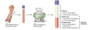
Blood consists of plasma (the liquid matrix) and formed elements (cells and cell fragments). The formed elements include erythrocytes (red blood cells), leukocytes (white blood cells), and platelets.
Plasma: Makes up about 55% of whole blood; contains water, proteins (albumin, globulins, fibrinogen), nutrients, gases, hormones, wastes, and inorganic ions.
Formed Elements:
Erythrocytes: Red blood cells, responsible for oxygen and carbon dioxide transport.
Leukocytes: White blood cells, involved in immune defense.
Platelets: Cell fragments essential for blood clotting.
Physical Characteristics and Volume
Blood is a viscous, opaque fluid with a metallic taste. Its viscosity is about five times greater than water, mainly due to erythrocytes. The color varies with oxygen content: bright red when oxygen-rich, dark red when oxygen-poor. Blood pH ranges from 7.35 to 7.45, and it constitutes about 8% of body weight (average volume: 5–6 L in males, 4–5 L in females).
Blood Plasma
Composition of Plasma
Plasma is a straw-colored, sticky fluid composed mostly of water and over 100 dissolved solutes. Plasma proteins are the most abundant solutes by weight, primarily produced by the liver (except antibodies and hormones).
Albumin: Most abundant plasma protein; functions as a blood buffer, carrier for certain molecules, and contributor to plasma osmotic pressure.
Other Solutes: Nutrients, gases, hormones, wastes, and inorganic ions.
Formed Elements
General Features
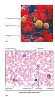
The formed elements include erythrocytes, leukocytes, and platelets. Erythrocytes and platelets are not true cells (erythrocytes lack nuclei and most organelles; platelets are cell fragments). Most formed elements survive only a few days and are replaced by stem cells in red bone marrow.
Erythrocytes (Red Blood Cells)
Structural Characteristics
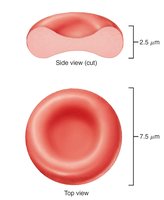
Erythrocytes are small, biconcave cells lacking a nucleus and most organelles. They are filled with hemoglobin for gas transport and have a flexible cytoskeleton allowing passage through narrow capillaries. Their structure maximizes surface area for gas exchange and minimizes diffusion distance.
Biconcave Shape: Provides a large surface area to volume ratio.
Absence of Mitochondria: Prevents consumption of transported oxygen; ATP is produced anaerobically.
Functions of Erythrocytes
RBCs are dedicated to respiratory gas transport. Hemoglobin binds reversibly with oxygen and carbon dioxide.
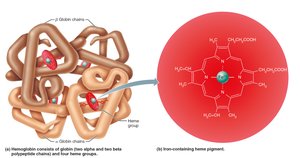
Hemoglobin: Consists of four globin polypeptide chains (two alpha, two beta) and four heme groups, each with a central iron ion that binds one oxygen molecule.
Oxygen Transport: Each hemoglobin molecule can carry four oxygen molecules; each RBC contains millions of hemoglobin molecules.
Color: Oxygen-bound hemoglobin (oxyhemoglobin) is bright red; deoxygenated hemoglobin (deoxyhemoglobin) is dark red.
Carbon Dioxide Transport: 20% of CO2 transported in blood binds to hemoglobin, forming carbaminohemoglobin.
Production of Erythrocytes (Erythropoiesis)
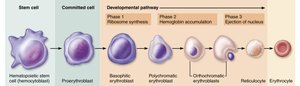
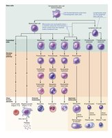
Hematopoiesis occurs in red bone marrow, where hematopoietic stem cells (hemocytoblasts) give rise to all formed elements. Erythropoiesis is the process of erythrocyte production, involving several developmental stages.
Stages: Hematopoietic stem cell → myeloid stem cell → proerythroblast → basophilic erythroblast → polychromatic erythroblast → orthochromatic erythroblast → reticulocyte → erythrocyte.
Reticulocyte Count: Used to estimate the rate of RBC formation.
Regulation and Requirements of Erythropoiesis
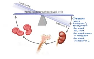
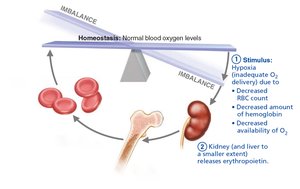
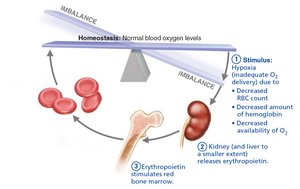
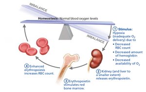
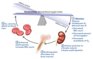
The number of circulating RBCs is tightly regulated to maintain homeostasis. Erythropoiesis is controlled hormonally, primarily by erythropoietin (EPO), and depends on adequate dietary nutrients.
Erythropoietin (EPO): Produced by kidneys in response to hypoxia; stimulates RBC production.
Dietary Requirements: Amino acids, lipids, carbohydrates, vitamin B12, folic acid, and iron are essential for erythropoiesis.
Fate and Destruction of Erythrocytes
RBCs have a lifespan of 100–120 days. They are removed from circulation by macrophages in the spleen, liver, and bone marrow. Hemoglobin is broken down into heme, iron, and globin; heme is converted to bilirubin, iron is stored or reused, and globin is metabolized into amino acids.
Leukocytes (White Blood Cells)
General Structure and Function
Leukocytes are complete cells with nuclei and organelles. They defend the body against pathogens and are capable of leaving the bloodstream to enter tissues (diapedesis).
Leukocytosis: Elevated WBC count, often a response to infection.
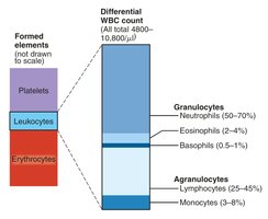
Classification:
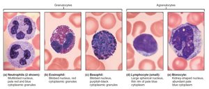
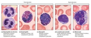
Granulocytes: Neutrophils, eosinophils, basophils (contain cytoplasmic granules).
Agranulocytes: Lymphocytes, monocytes (lack visible granules).
Granulocytes
Neutrophils: Most numerous; phagocytic; kill bacteria with defensins and oxidizing substances.
Eosinophils: Attack parasitic worms; modulate immune responses; involved in allergies and asthma.
Basophils: Release histamine; involved in inflammatory responses.
Agranulocytes
Lymphocytes: Second most numerous; T cells act against virus-infected and tumor cells; B cells produce antibodies.
Monocytes: Largest WBCs; differentiate into macrophages; active in phagocytosis and immune response activation.
Production and Life Span of Leukocytes
Leukopoiesis is stimulated by interleukins and colony-stimulating factors. Hematopoietic stem cells differentiate into myeloid or lymphoid stem cells, giving rise to various leukocytes.
Platelets
Structure and Function
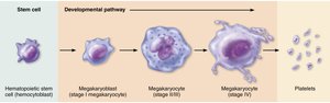
Platelets are cell fragments derived from megakaryocytes. They contain granules with chemicals essential for clotting and form temporary plugs to seal vessel breaks.
Formation: Regulated by thrombopoietin; megakaryocytes fragment in bone marrow and lungs to produce platelets.
Normal Concentration: 150,000–400,000 platelets/mL.
Hemostasis
Overview
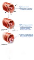
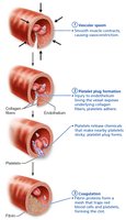
Hemostasis is the process that prevents blood loss following vessel injury. It involves three steps: vascular spasm, platelet plug formation, and coagulation.
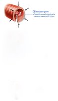
Step 1: Vascular Spasm: Vasoconstriction reduces blood loss.
Step 2: Platelet Plug Formation: Platelets adhere to exposed collagen, become activated, and aggregate.
Step 3: Coagulation: Reinforces the platelet plug with a fibrin mesh.
Coagulation Pathways
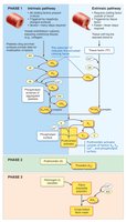
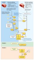
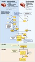
Coagulation involves a cascade of reactions leading to the formation of a fibrin clot. There are intrinsic and extrinsic pathways, both converging to activate prothrombin.
Intrinsic Pathway: Triggered by factors within the blood.
Extrinsic Pathway: Triggered by tissue factor from outside the blood.
Common Pathway: Prothrombin is converted to thrombin, which then converts fibrinogen to fibrin.

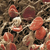
Fibrin Mesh and Clot Formation
The fibrin mesh traps erythrocytes and platelets, forming a stable blood clot. This process is essential for sealing larger vessel breaks and preventing excessive blood loss.
Summary Table: Composition of Blood
Component | Main Function | Relative Abundance |
|---|---|---|
Plasma | Transport, regulation, protection | ~55% |
Erythrocytes | O2 and CO2 transport | ~45% |
Leukocytes | Immune defense | <1% |
Platelets | Clotting | <1% |
Key Equations
Hemoglobin Oxygen Binding:
Hematocrit Calculation:
Additional info:
Vitamin K is required for synthesis of several clotting factors.
Reticulocyte counts are clinically used to assess bone marrow function.
Leukocyte classification mnemonic: "Never Let Monkeys Eat Bananas" (Neutrophils, Lymphocytes, Monocytes, Eosinophils, Basophils).
