 Back
Blood Vessels and Circulation: Structure, Function, and Regulation
Study Guide - Smart Notes
 Tailored notes based on your materials, expanded with key definitions, examples, and context.
Blood Vessels: Overview and Classification
Types of Blood Vessels
The vascular system is composed of several types of blood vessels, each with distinct structural and functional characteristics. These vessels form a closed circuit for blood flow throughout the body.
Arteries: Carry blood away from the heart's ventricles to tissues.
Arterioles: Small branches of arteries that lead to capillaries; regulate blood flow and pressure.
Capillaries: Microscopic vessels where exchange of gases, nutrients, and wastes occurs between blood and tissues.
Venules: Collect blood from capillaries and transport it to veins.
Veins: Return blood to the atria of the heart; often contain valves to prevent backflow.
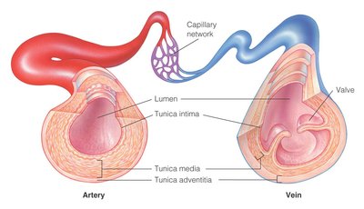
Structure of Blood Vessel Walls
Layers of Blood Vessels
Most arteries and veins share a similar three-layered structure, but the thickness and composition of these layers vary according to vessel type and function.
Tunica Externa (Adventitia): Outermost connective tissue layer; provides support and anchors vessels to surrounding tissues.
Tunica Media: Middle layer composed of smooth muscle and elastic fibers; responsible for vasoconstriction and vasodilation.
Tunica Interna (Intima): Innermost layer of squamous endothelium; minimizes friction between blood and vessel wall.
Veins have thinner walls and larger lumens than arteries, and many contain valves to prevent backflow due to lower pressure.
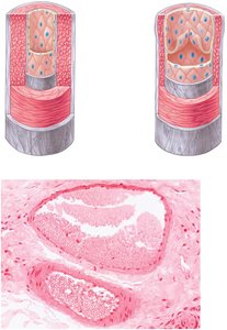
Arteries and Arterioles
Functional Types and Roles
Arteries are classified based on their size and function:
Elastic (Conducting) Arteries: Largest arteries; act as pressure reservoirs (e.g., aorta).
Muscular (Distributing) Arteries: Medium-sized; distribute blood to organs.
Arterioles: Smallest arteries; regulate blood flow into capillaries by changing diameter (vasoconstriction/vasodilation).
Arterioles are known as resistance vessels because their contraction or relaxation significantly affects blood pressure and flow.
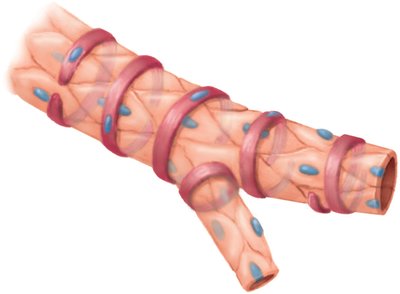
Capillaries
Structure and Function
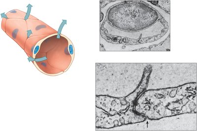
Capillaries are the smallest blood vessels, consisting of a single layer of endothelial cells. They connect arterioles to venules and are the primary sites for exchange between blood and tissues.
Precapillary sphincters regulate blood flow into capillary beds.
Capillaries are semi-permeable, allowing selective exchange of substances.
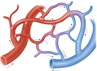
Capillary Exchange Mechanisms
Exchange across capillary walls occurs via diffusion, filtration, and osmosis. The structure of capillary walls facilitates efficient movement of gases, nutrients, and wastes.
Venules and Veins
Structure and Function
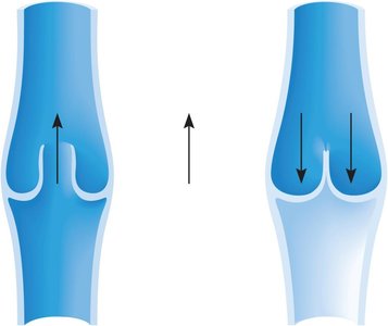
Venules collect blood from capillaries and merge to form veins. Veins have thinner walls, larger lumens, and lower pressure compared to arteries. Many veins, especially in the limbs, contain valves to prevent backflow of blood.
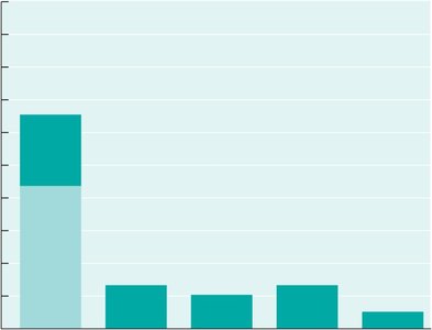
Veins serve as blood reservoirs, containing about 65% of the body's blood volume.
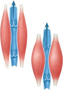
Venous return is aided by skeletal muscle contraction, respiratory movements, and venoconstriction.
Distribution of Blood in the Vascular System
Blood Volume Distribution
The majority of blood volume is found in the systemic veins and venules, reflecting their role as blood reservoirs.
Blood Pressure and Its Regulation
Blood Pressure Definitions
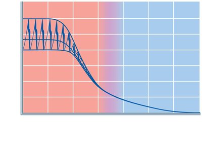
Blood pressure is the force exerted by circulating blood on the walls of blood vessels. It is highest in the arteries and decreases through the capillaries and veins.
Systolic Pressure: Maximum pressure during ventricular contraction.
Diastolic Pressure: Minimum pressure during ventricular relaxation.
Mean Arterial Pressure (MAP): Average pressure in the arteries during one cardiac cycle.
MAP can be calculated as:
or
where SBP = systolic blood pressure, DBP = diastolic blood pressure.
Baroreceptor Reflex and Blood Pressure Homeostasis
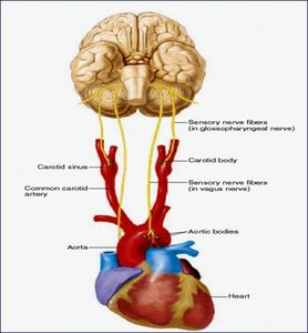
Baroreceptors are stretch-sensitive mechanoreceptors located in the aortic arch and carotid sinuses. They detect changes in blood pressure and initiate reflexes to maintain homeostasis via the autonomic nervous system.
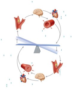
Increased blood pressure stimulates baroreceptors, leading to decreased heart rate and vasodilation.
Decreased blood pressure reduces baroreceptor firing, resulting in increased heart rate and vasoconstriction.
Factors Influencing Blood Pressure
Several factors affect arterial blood pressure:
Blood volume
Heart rate
Stroke volume
Blood viscosity
Peripheral resistance
Blood pressure is regulated by neural, hormonal, and autoregulatory mechanisms.
Hormonal Regulation of Blood Pressure
Key hormones involved in blood pressure regulation include:
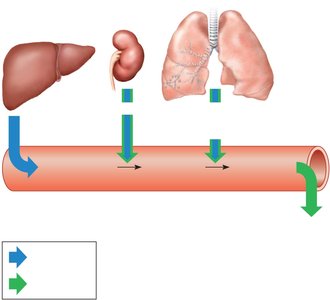
Antidiuretic Hormone (ADH): Increases water reabsorption in kidneys, raising blood volume and pressure.
Aldosterone: Increases sodium (and water) reabsorption in kidneys, increasing blood volume and pressure.
Renin-Angiotensin System: Renin release leads to formation of angiotensin II, which causes vasoconstriction and stimulates ADH and aldosterone release.
Atrial Natriuretic Peptide (ANP): Released by the heart in response to high blood pressure; increases sodium and water excretion, lowering blood volume and pressure.
Venous Blood Flow and Central Venous Pressure
Mechanisms of Venous Return
Venous return to the heart is facilitated by:
Skeletal muscle pump
Respiratory pump
Venoconstriction
One-way venous valves
Central Venous Pressure (CVP)
CVP reflects the amount of blood returning to the heart and the heart's ability to pump it into the arterial system. Elevated CVP can lead to peripheral edema.
Systemic Circulation: Major Arteries and Veins
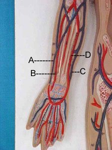
Major Arteries of the Systemic Circuit
The systemic circuit delivers oxygenated blood from the left ventricle to all body tissues (except the lungs) and returns deoxygenated blood to the right atrium.
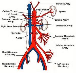
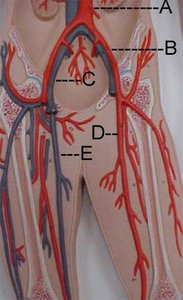
Aorta: Main artery leaving the left ventricle; branches supply the head, neck, upper limbs, and trunk.
Major branches: Brachiocephalic, left common carotid, left subclavian, celiac trunk, mesenteric arteries, renal arteries, common iliac arteries, femoral arteries, and others.
Major Veins of the Systemic Circuit
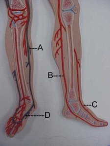
Veins return deoxygenated blood to the heart. Major veins include the superior and inferior vena cava, jugular veins, subclavian veins, brachiocephalic veins, femoral veins, and the great saphenous vein.
Hepatic Portal System
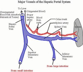
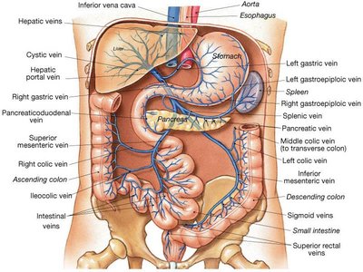
The hepatic portal vein collects blood from the digestive organs, spleen, and pancreas, directing it to the liver for processing before it enters the systemic circulation.
Vessel Dynamics and Blood Flow Equations
Key Equations
Cardiac Output (CO): The volume of blood ejected by the ventricles per minute.
Blood Pressure (BP): Determined by cardiac output and peripheral resistance.
Vessel radius, length, and blood viscosity all influence resistance and thus blood pressure.
Additional info: Increased vessel radius decreases resistance and blood pressure, while increased vessel length or viscosity increases resistance and blood pressure.
