 Back
BackBlood Vessels: Structure, Function, and Circulatory Pathways
Study Guide - Smart Notes
 Tailored notes based on your materials, expanded with key definitions, examples, and context.
Blood Vessels: Structure and Function
Overview of Blood Vessels
Blood vessels are essential components of the cardiovascular system, responsible for transporting blood throughout the body. They include arteries, veins, and capillaries, each with distinct structural and functional characteristics.
Arteries: Carry blood away from the heart to tissues.
Veins: Return blood to the heart from tissues.
Capillaries: Connect arteries and veins; site of nutrient and gas exchange.
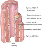
Microscopic Anatomy of Blood Vessels
Blood vessels are composed of three main layers (tunics):
Tunica intima (interna): Innermost layer; consists of endothelium (simple squamous epithelium) and a basement membrane. Provides a smooth surface for blood flow.
Tunica media: Middle layer; composed of smooth muscle and elastic fibers. Responsible for vasoconstriction and vasodilation, regulated by the sympathetic nervous system.
Tunica externa (adventitia): Outermost layer; made of connective tissue with collagen and elastic fibers. Provides structural support and protection.
Classification of Arteries
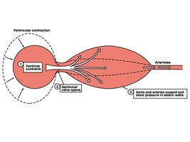
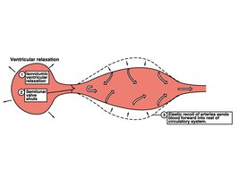
Elastic arteries (conducting arteries): Large vessels near the heart (e.g., aorta). Contain abundant elastic tissue to withstand and smooth out pressure fluctuations from the heart.
Muscular arteries (distributing arteries): Medium-sized; deliver blood to specific body regions. More smooth muscle, less elastic tissue.
Arterioles: Smallest arteries; regulate blood flow into capillary beds by changing diameter.
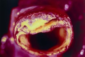
Arteriosclerosis and Atherosclerosis
Arteriosclerosis: General term for the thickening and loss of elasticity of arterial walls.
Atherosclerosis: A specific type of arteriosclerosis involving lipid deposits (plaques) in the arterial wall, leading to narrowed and stiffened arteries.
Aneurysms
An aneurysm is a localized dilation or ballooning of a blood vessel wall, often due to weakness in the vessel. Common sites include the abdominal aorta and cerebral arteries. Risk factors include atherosclerosis and hypertension.

Capillaries and Capillary Beds
Structure and Function of Capillaries
Capillaries are the smallest blood vessels, consisting only of endothelium and a basement membrane. They are the primary site for exchange of gases, nutrients, and wastes between blood and tissues.
Diameter: ~8-10 μm (just large enough for red blood cells to pass through in single file)
Provide direct access to nearly every cell in the body
Types of Capillaries
Continuous capillaries: Most common; found in skin and muscles. Endothelial cells joined by tight junctions with small intercellular clefts for limited permeability.
Fenestrated capillaries: Have pores (fenestrations) in endothelial cells; found in areas of active filtration (kidneys, small intestine, endocrine glands).
Sinusoidal capillaries (sinusoids): Large, irregular lumens and incomplete basement membranes; found in liver, bone marrow, spleen, and adrenal medulla. Highly permeable to allow passage of large molecules and cells.
Capillary Beds and Blood Flow Regulation
Capillary beds consist of networks of capillaries supplied by arterioles and drained by venules. Blood flow through capillary beds is regulated by precapillary sphincters, which open or close in response to local chemical signals and neural input.
Veins and Venous Return
Structure and Function of Veins
Veins carry blood back to the heart and act as blood reservoirs, containing up to 65% of the body's blood volume. They have thinner walls and larger lumens than arteries, and lower blood pressure.
Valves in veins prevent backflow of blood, especially in limbs.
Venous return is aided by the muscular pump (skeletal muscle contraction), respiratory pump (pressure changes during breathing), and sympathetic venoconstriction.
Venous Disorders
Varicose veins: Result from leaky venous valves, leading to blood pooling and vein dilation.
Hemorrhoids: Varicosities in the veins of the anal canal due to increased venous pressure.
Vascular Anastomoses
Vascular anastomoses are interconnections between blood vessels that provide alternate pathways for blood flow. They are common in joints, abdominal organs, the brain, and the heart. Arteriovenous anastomoses allow blood to bypass capillary beds.
Blood Flow, Blood Pressure, and Regulation
Blood Flow and Blood Pressure
Blood flow: Volume of blood flowing through a vessel, organ, or the entire circulation in a given period (mL/min).
Blood pressure (BP): Force per unit area exerted on a vessel wall by the contained blood (measured in mmHg).
BP is highest in the aorta and lowest in the venae cavae.
Key Blood Pressure Terms
Systolic pressure: Pressure during ventricular contraction (~120 mmHg).
Diastolic pressure: Pressure during ventricular relaxation (~70-80 mmHg).
Pulse pressure: Difference between systolic and diastolic pressure.
Mean arterial pressure (MAP): Average pressure in arteries during one cardiac cycle. Calculated as
Factors Affecting Blood Flow and Pressure
Peripheral resistance: Opposition to flow, mainly due to vessel diameter, blood viscosity, and vessel length.
Cardiac output: Volume of blood pumped by the heart per minute.
Blood volume: Total amount of blood in the vascular system.
Regulation of Blood Pressure
Short-term regulation: Neural and hormonal mechanisms (baroreceptors, chemoreceptors, sympathetic/parasympathetic nervous system, hormones such as epinephrine, norepinephrine, angiotensin II, ADH, and ANP).
Long-term regulation: Renal mechanisms that adjust blood volume via water and salt balance.
Capillary Exchange Mechanisms
Diffusion
Movement of molecules down their concentration gradients through capillary walls (e.g., O2, CO2, glucose, amino acids).
Bulk Flow
Movement of fluid and solutes due to pressure gradients. Involves hydrostatic and osmotic pressures:
Hydrostatic pressure (HP): Pushes fluid out of capillaries (filtration).
Osmotic pressure (OP): Draws fluid into capillaries (reabsorption).
At the arterial end, HP > OP, so fluid leaves the capillary. At the venous end, OP > HP, so fluid enters the capillary.
Circulatory Pathways
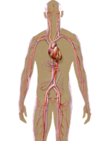
Systemic Circulation
Delivers oxygenated blood from the heart to body tissues and returns deoxygenated blood to the heart.
Pulmonary Circulation
Delivers deoxygenated blood from the heart to the lungs for gas exchange and returns oxygenated blood to the heart.
Hepatic Portal Circulation
Collects nutrient-rich blood from the gastrointestinal tract and routes it to the liver for processing before entering general circulation.
Blood Pressure Disorders
Hypertension: Sustained arterial pressure of 140/90 mmHg or higher. Major risk factor for heart disease, stroke, and kidney failure.
Hypotension: Abnormally low blood pressure (typically below 90/60 mmHg). May cause dizziness or fainting.
Circulatory shock: Inadequate blood flow to meet tissue needs. Types include hypovolemic, vascular, neurogenic, and cardiogenic shock.
