 Back
BackBlood Vessels: Structure, Function, and Major Pathways
Study Guide - Smart Notes
 Tailored notes based on your materials, expanded with key definitions, examples, and context.
Blood Vessel Structure and Function
Generalized Structure of Blood Vessels
The circulatory system is composed of arteries, veins, and capillaries, each with distinct structural features that support their functions in transporting blood throughout the body.
Arteries have thick walls with three layers: tunica intima (innermost), tunica media (middle, smooth muscle and elastic fibers), and tunica externa (outer, collagen fibers). They carry blood away from the heart under high pressure.
Veins have thinner walls, larger lumens, and often contain valves to prevent backflow. They return blood to the heart under lower pressure.
Capillaries are composed of a single layer of endothelial cells and a basement membrane, allowing for efficient exchange of gases, nutrients, and wastes between blood and tissues.
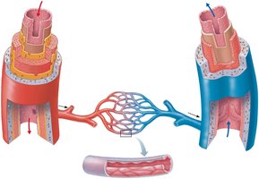
Summary of Blood Vessel Anatomy and Physiology
Blood vessels are classified based on their structure and function. The following table summarizes the main types of blood vessels, their characteristics, and their roles in circulation.
Type of Vessel | Description | Average Lumen Diameter | Average Wall Thickness | Function |
|---|---|---|---|---|
Elastic (conducting) arteries | Largest arteries; contain more elastic tissue than other vessels | 1.5 cm | 1.0 mm | Act as pressure reservoirs; expand and recoil for continuous blood flow |
Muscular (distributing) arteries | Medium-sized arteries; distribute blood to specific organs | 0.6 cm | 1.0 mm | Deliver blood to body organs; active in vasoconstriction |
Arterioles | Smallest arteries; control flow into capillary beds | 37 µm | 6.0 µm | Regulate blood flow and pressure |
Capillaries | Smallest blood vessels; walls are one cell thick | 9 µm | 0.5 µm | Permit exchange of nutrients, gases, and wastes |
Venules | Smallest veins; drain capillary beds | 20 µm | 1.0 µm | Collect blood from capillaries and transport to veins |
Veins | Carry blood toward the heart; thinner walls and larger lumens than arteries | 5.0 mm | 0.5 mm | Low-pressure vessels; valves prevent backflow |
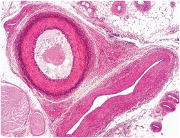
Histology of Arteries and Veins
Microscopic examination reveals differences in the wall structure of arteries and veins. Arteries have a thicker tunica media, while veins have a larger lumen and thinner wall. These differences reflect their functional roles in the circulatory system.
Arteries: Thick tunica media for withstanding high pressure.
Veins: Thin walls and valves to accommodate low-pressure blood flow and prevent backflow.
Major Arterial Pathways
Systemic Arterial Circulation
The systemic arterial system delivers oxygenated blood from the left ventricle of the heart to all body tissues. The aorta is the main artery, branching into major arteries that supply the head, neck, upper limbs, thorax, abdomen, pelvis, and lower limbs.
Aorta: The largest artery, divided into ascending aorta, aortic arch, thoracic aorta, and abdominal aorta.
Major branches: Include the brachiocephalic trunk, common carotid arteries, subclavian arteries, and branches to the thoracic and abdominal organs.
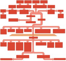
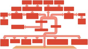
Arteries of the Head, Neck, and Brain
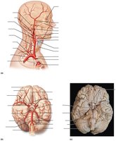
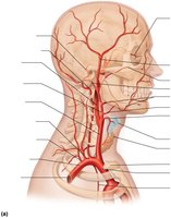
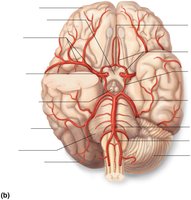
The head and neck receive blood from the common carotid and vertebral arteries. The brain is supplied by the internal carotid and vertebral arteries, which form the cerebral arterial circle (Circle of Willis) for collateral circulation.
External carotid artery: Supplies face and scalp.
Internal carotid artery: Supplies brain.
Vertebral artery: Supplies posterior brain regions.
Arteries of the Upper Limb and Thorax
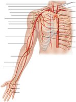
The upper limb is supplied by branches of the subclavian artery, which continues as the axillary, brachial, radial, and ulnar arteries. The thoracic aorta gives off branches to the thoracic wall and organs.
Axillary artery: Supplies the shoulder and upper arm.
Brachial artery: Main artery of the arm.
Radial and ulnar arteries: Supply the forearm and hand.
Arteries of the Abdomen
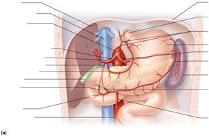
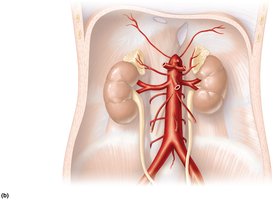
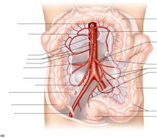
The abdominal aorta supplies the abdominal organs through several major branches, including the celiac trunk, superior mesenteric artery, renal arteries, and inferior mesenteric artery.
Celiac trunk: Supplies the liver, stomach, spleen, and upper duodenum.
Superior mesenteric artery: Supplies the small intestine and part of the large intestine.
Renal arteries: Supply the kidneys.
Inferior mesenteric artery: Supplies the distal large intestine.
Arteries of the Pelvis and Lower Limb
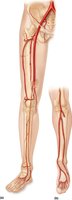
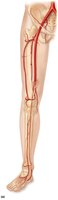
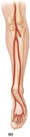
The common iliac arteries branch into internal and external iliac arteries, supplying the pelvis and lower limbs. The femoral artery continues as the popliteal, anterior tibial, posterior tibial, and fibular arteries.
Femoral artery: Main artery of the thigh.
Popliteal artery: Behind the knee.
Anterior and posterior tibial arteries: Supply the leg and foot.
Major Venous Pathways
Systemic Venous Circulation
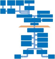
Veins return deoxygenated blood from the body to the right atrium of the heart. The superior and inferior venae cavae are the main veins, receiving blood from the upper and lower body, respectively.
Superior vena cava: Drains blood from the head, neck, upper limbs, and thorax.
Inferior vena cava: Drains blood from the abdomen, pelvis, and lower limbs.
Veins of the Pelvis and Lower Limb
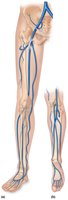
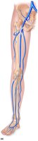
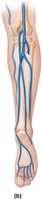
The veins of the lower limb include the great saphenous vein (superficial), femoral vein (deep), and their tributaries. These veins drain into the external and common iliac veins, which join to form the inferior vena cava.
Great saphenous vein: Longest vein in the body; often used in coronary bypass surgery.
Femoral vein: Main deep vein of the thigh.
Venous Drainage of the Abdomen
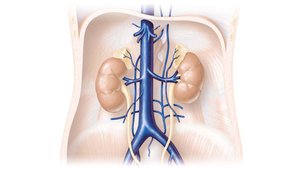
Abdominal organs not drained by the hepatic portal vein return blood directly to the inferior vena cava via the renal, suprarenal, gonadal, and lumbar veins.
Venous Drainage of the Head, Neck, and Brain
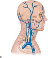
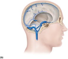
Venous blood from the brain drains into dural venous sinuses, which empty into the internal jugular veins. The external jugular and vertebral veins drain superficial structures and the cervical spinal cord.
Veins of the Thorax and Upper Limb
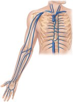
The veins of the upper limb include the cephalic, basilic, and median cubital veins (superficial), and the brachial, axillary, and subclavian veins (deep). The azygos system drains the thoracic wall.
Special Circulatory Pathways
Pulmonary Circulation
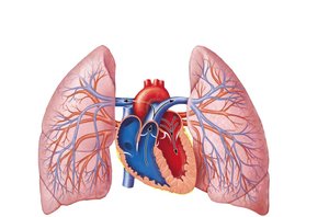
Pulmonary circulation carries deoxygenated blood from the right ventricle to the lungs via the pulmonary arteries and returns oxygenated blood to the left atrium via the pulmonary veins.
Fetal and Newborn Circulation
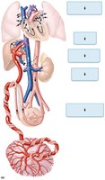
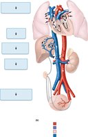
Fetal circulation includes unique structures such as the ductus arteriosus, foramen ovale, and ductus venosus, which allow blood to bypass the nonfunctional fetal lungs and liver. After birth, these structures close or become ligaments.
Ductus arteriosus: Becomes ligamentum arteriosum.
Foramen ovale: Becomes fossa ovalis.
Ductus venosus: Becomes ligamentum venosum.
Hepatic Portal Circulation
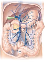
The hepatic portal system carries nutrient-rich blood from the digestive organs to the liver for processing before it enters the systemic circulation. Major vessels include the hepatic portal vein, superior mesenteric vein, and splenic vein.
Key Arteries and Veins to Know for Labelling
Arteries: Aorta, common carotid, subclavian, axillary, brachial, radial, ulnar, celiac, renal, inferior mesenteric, common iliac, internal iliac, external iliac, femoral, popliteal, anterior tibial, posterior tibial
Veins: Internal jugular, subclavian, brachiocephalic, superior vena cava, axillary, cephalic, basilic, brachial, median cubital, hepatic, renal, inferior vena cava, common iliac, internal iliac, external iliac, great saphenous, femoral, dorsal venous arch
Additional info: Understanding the structure and function of blood vessels is essential for interpreting clinical signs, performing procedures, and understanding the pathophysiology of cardiovascular diseases.
