 Back
BackBlood Vessels: Structure, Function, and Regulation
Study Guide - Smart Notes
 Tailored notes based on your materials, expanded with key definitions, examples, and context.
Blood Vessels: Overview
Types of Blood Vessels
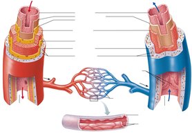
Blood vessels are essential components of the circulatory system, responsible for transporting blood throughout the body. The three main types are arteries, veins, and capillaries.
Arteries: Carry blood away from the heart. They are involved in both systemic and pulmonary circulation.
Veins: Carry blood toward the heart. Also participate in systemic and pulmonary circulation.
Capillaries: Serve as the site of gas and nutrient exchange between blood and tissues.
 Learn
Learn
Circulation Pathways
Blood flows from the heart to the lungs and tissues, then returns to the heart, completing the circuit of systemic and pulmonary circulation.
Structure of Blood Vessel Walls
Layers (Tunics) of Blood Vessels
Most blood vessels (except capillaries) have three distinct layers:
Tunica interna (intima): The innermost layer, composed of endothelium and underlying connective tissue. It lines the lumen and is in direct contact with blood.
Tunica media: The middle layer, consisting of circular smooth muscle and elastic fibers. It maintains blood pressure and controls vessel diameter.
Tunica externa (adventitia): The outermost layer, made mostly of collagen fibers. It anchors and reinforces vessels. Large vessels contain vasa vasorum, a network of small vessels, nerves, and lymphatics supplying the outer wall.
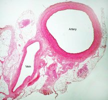
Arteries have a thicker tunica media and more elastin, while veins have a thicker tunica externa. Vessel characteristics vary in length, diameter, wall thickness, and tissue composition.
Classification of Blood Vessels
Arteries
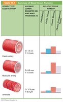
Elastic arteries (conducting vessels): Large vessels near the heart (e.g., aorta). They have large lumens, low resistance, and abundant elastin in the tunica media.
Muscular arteries (distributing vessels): Medium-sized arteries (e.g., brachial, radial). Their tunica media contains more smooth muscle and less elastin, regulated by the sympathetic nervous system.
Arterioles (resistance vessels): Smallest arteries, regulate blood flow into capillary beds by changing diameter in response to neural, hormonal, and chemical stimuli.
Capillaries
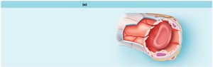
Capillaries are the smallest blood vessels, consisting only of the tunica interna. They are the primary site for exchange of gases, nutrients, wastes, and hormones.
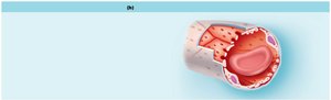
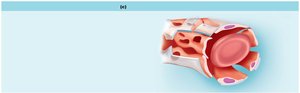
Continuous capillaries: Least permeable and most common, found in skin, muscles, lungs, and CNS. Associated with pericytes and tight junctions.
Fenestrated capillaries: Have large pores (fenestrations) that increase permeability. Found in areas of active filtration (kidney), absorption (small intestine), and endocrine secretion.
Sinusoidal capillaries: Most permeable, found in liver, bone marrow, spleen, and adrenal medulla. Have large intercellular clefts, incomplete basement membranes, and allow passage of large molecules and cells.
Capillary Beds and Microcirculation
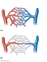
Microcirculation refers to the flow of blood from arterioles to venules through capillaries. Precapillary sphincters regulate blood flow into capillary beds, adjusting to local chemical conditions.
Portal Systems
A portal system consists of two capillary networks joined by a vein, such as the hepatic portal circulation.
Veins and Venules
Venules: Smallest veins, very porous, allow movement of WBCs and fluid.
Veins (capacitance vessels): Formed by joining venules, have thinner walls and larger lumens than arteries. The thickest layer is tunica externa. Veins hold about 65% of the blood supply and require mechanisms (valves, muscular and respiratory pumps) to return blood to the heart.
Blood Flow, Pressure, and Resistance
Blood Flow (BF)
Blood flow is the volume of blood moving through vessels, organs, or the entire circulation per unit time. It is equivalent to cardiac output (CO).
CO = HR x SV (Heart Rate x Stroke Volume)
BF = DP/R (Blood Flow = Pressure Difference / Resistance)
Pressure and Resistance
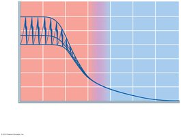
Pressure: Generated by ventricular contraction, measured in mm Hg. Systolic (120 mmHg) and diastolic (80 mmHg) are typical values.
Pulse Pressure: Difference between systolic and diastolic pressure (e.g., 40 mmHg).
Mean Arterial Pressure (MAP): Average pressure in the arteries, necessary for organ perfusion.
Resistance to Blood Flow
Blood viscosity: Thickness of blood, determined by ratio of cells to plasma.
Vessel length: Longer vessels increase resistance.
Lumen diameter: Most important factor; smaller diameter increases resistance. (Poiseuille’s Law)
Mechanisms Assisting Venous Return
Respiratory Pump
Pressure changes during breathing help move blood toward the heart.
Muscular Pump
Contraction of skeletal muscles "milks" blood upward in veins.
Venous Valves
Valves prevent backflow of blood, ensuring one-way movement toward the heart.
Regulation of Blood Pressure and Blood Flow
Neural Mechanisms
Cardiovascular center (medulla): Includes cardioinhibitory (parasympathetic), cardioacceleratory (sympathetic), and vasomotor (sympathetic) centers.
Baroreceptors: Respond to pressure changes, located in aortic arch and carotid sinus.
Chemoreceptors: Monitor O2, CO2, and pH, located near baroreceptors.
Higher brain centers: Cerebral cortex, limbic system, hypothalamus can modify cardiovascular responses.
Hormonal Control
ADH: Increases water reabsorption, raising blood pressure.
Epinephrine/Norepinephrine: Increase heart rate and vasoconstriction.
Angiotensin II/Aldosterone: Increase blood pressure via vasoconstriction and sodium retention.
Atrial natriuretic peptide (ANP): Decreases blood pressure by promoting sodium and water excretion.
Renal Regulation
Kidneys regulate blood pressure by controlling blood volume directly (filtration and urine production) and indirectly (renin-angiotensin-aldosterone mechanism).
Autoregulation of Blood Flow
Metabolic and Myogenic Controls
Metabolic control: Decreased O2, increased CO2, H+, K+, and nitric oxide cause vasodilation.
Myogenic control: Vessel stretch causes reflexive vasoconstriction; low pressure causes vasodilation.
Long-term: Persistent increased demand leads to angiogenesis (new vessel formation).
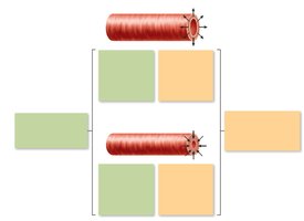
Capillary Exchange and Transport Mechanisms
Mechanisms of Capillary Transport
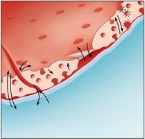
Lipid-soluble substances: Diffuse through endothelial membranes.
Water-soluble substances: Move through intercellular clefts or fenestrations.
Vesicular transport: Large substances transported via vesicles.
Bulk Flow of Fluid in Capillaries
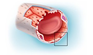
Bulk flow is the passive movement of fluid down a pressure gradient, determining fluid volumes in body compartments. It depends on hydrostatic and osmotic pressures.
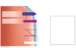
Hydrostatic pressure: Pushes water out of capillaries.
Osmotic pressure: Pulls water into capillaries, mainly due to plasma proteins.
Filtration and Reabsorption
Filtration: Movement of fluid out of capillaries at the arterial end.
Reabsorption: Movement of fluid into capillaries at the venous end.
Net Filtration Pressure (NFP)
NFP determines the direction of fluid movement:
Positive NFP: Filtration (fluid leaves capillary)
Negative NFP: Reabsorption (fluid enters capillary)
Summary Table: Blood Vessel Anatomy
Vessel Type | Diameter | Wall Thickness | Relative Tissue Makeup |
|---|---|---|---|
Elastic artery | 1.5 cm | 1.0 mm | High elastin, moderate smooth muscle, constant endothelium |
Muscular artery | 0.6 cm | 1.0 mm | High smooth muscle, less elastin, constant endothelium |
Arteriole | 37 μm | 6.5 μm | Mostly smooth muscle, minimal elastin, constant endothelium |
Practice Questions and Applications
Which vessels carry blood away from the heart? Arteries
Name the innermost blood vessel layer: Tunica interna (intima)
What is the main determinant of resistance? Lumen diameter
How does increasing resistance affect blood flow? Decreases blood flow
What is the formula for mean arterial pressure?
What is the formula for net filtration pressure?
Example: If HPc = 30 mmHg, OPc = 22 mmHg, HPif = 0 mmHg, OPif = 1 mmHg, then mmHg. Positive NFP means net filtration (fluid leaves capillaries).
