Back
BackBone Tissue: Structure, Function, and Development
Study Guide - Smart Notes
 Tailored notes based on your materials, expanded with key definitions, examples, and context.
Bone Tissue
Functions of the Skeletal System
The skeletal system performs several essential functions that are critical for maintaining homeostasis and supporting life.
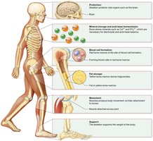
Protection: Bones protect vital organs such as the brain (skull), heart, and lungs (rib cage).
Mineral Storage and Acid-Base Homeostasis: Bones store minerals like calcium (Ca2+) and phosphate (PO43-), releasing them into the blood as needed to maintain mineral balance and buffer blood pH.
Blood Cell Formation: Hematopoiesis occurs in red bone marrow, producing red blood cells, white blood cells, and platelets.
Fat Storage: Yellow bone marrow stores triglycerides, serving as an energy reserve.
Movement: Bones act as levers for muscles, enabling body movement.
Support: The skeleton provides structural support for the body and anchors soft tissues.
Classification of Bones by Shape
Bones are classified according to their shapes, which relate to their functions and locations in the body.
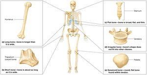
Long Bones: Longer than they are wide (e.g., humerus, femur).
Short Bones: About as long as they are wide (e.g., carpals, tarsals).
Flat Bones: Thin, broad, and often curved (e.g., skull bones, sternum, ribs, pelvis).
Irregular Bones: Complex shapes that do not fit other categories (e.g., vertebrae).
Sesamoid Bones: Small bones embedded within tendons (e.g., patella).
Wormian (Sutural) Bones: Extra bone pieces within skull sutures.
Structure of Long Bones vs. Other Bone Types
Long bones have a unique structure compared to short, flat, irregular, and sesamoid bones.
Long Bones:
Diaphysis: Shaft of the bone, composed mainly of compact bone surrounding the medullary cavity.
Epiphyses: Expanded ends, containing spongy bone and red marrow.
Articular Cartilage: Hyaline cartilage covering joint surfaces for smooth movement.
Periosteum: Dense outer layer with an inner osteogenic layer; attached to bone by perforating (Sharpey’s) fibers.
Endosteum: Thin membrane lining the medullary cavity and trabeculae of spongy bone.
Epiphyseal Plate/Line: Site of bone growth in length (hyaline cartilage in children, ossified in adults).
Short, Flat, Irregular, and Sesamoid Bones:
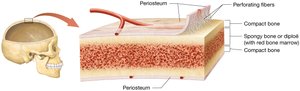
Composed of a thin outer layer of compact bone surrounding spongy bone (diploë in flat bones).
No diaphysis or epiphyses.
Contain bone marrow but lack a medullary cavity.
Red vs. Yellow Bone Marrow
Bone marrow exists in two forms, each with distinct functions and locations.
Red Marrow: Contains hematopoietic cells that produce blood cells; found in spongy bone of children and in select adult bones (e.g., pelvis, sternum).
Yellow Marrow: Consists mainly of adipocytes (fat cells); stores triglycerides and is found in the medullary cavity of long bones in adults.
Organic and Inorganic Components of the Bone Matrix
The extracellular matrix of bone is composed of both organic and inorganic materials, providing strength and flexibility.
Inorganic Matrix (65%): Primarily hydroxyapatite crystals (calcium and phosphate), along with bicarbonate, potassium, magnesium, and sodium. Provides hardness and resistance to compression.
Organic Matrix (35%): Known as osteoid; contains collagen fibers, proteoglycans, glycosaminoglycans (GAGs), and glycoproteins. Provides flexibility and tensile strength.
Bone Tissue Cells and Their Functions
Bone tissue contains several specialized cell types, each with a unique role in bone formation, maintenance, and resorption.
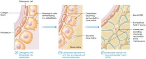
Osteogenic Cells: Stem cells found in the periosteum and endosteum; differentiate into osteoblasts.
Osteoblasts: Bone-building cells that secrete collagen and initiate matrix mineralization; become osteocytes when trapped in the matrix.
Osteocytes: Mature bone cells residing in lacunae; maintain bone matrix and signal for repair/remodeling.
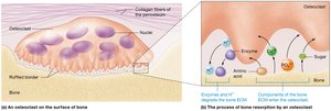
Osteoclasts: Large, multinucleated cells derived from monocytes; responsible for bone resorption by secreting acids and enzymes.
Compact vs. Spongy Bone Structure
Bone tissue is organized into two main types: compact and spongy bone, each with distinct structural features.
Compact Bone:
Dense and forms the outer layer of all bones.
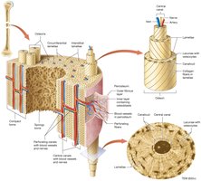
Composed of osteons (Haversian systems), which are cylindrical structures with concentric lamellae surrounding a central canal containing blood vessels and nerves.
Perforating (Volkmann’s) canals connect osteons and facilitate nutrient/waste exchange.
Spongy Bone (Cancellous Bone):
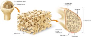
Located at the ends of long bones and inside flat, short, and irregular bones.
Composed of a network of trabeculae (bony struts) with spaces filled with bone marrow.
Lacks osteons; nutrients diffuse through canaliculi from blood vessels in the marrow spaces.
Bone Development and Growth
Bone Development: Primary vs. Secondary Bone
Bone formation (ossification or osteogenesis) occurs in two main stages and by two processes.
Primary (Woven) Bone: First bone formed during development or repair; irregular collagen arrangement, later replaced by secondary bone.
Secondary (Lamellar) Bone: Mature bone with organized collagen fibers and stronger structure.
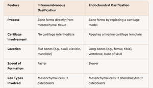
Intramembranous vs. Endochondral Ossification
There are two mechanisms of bone formation, each with distinct features and locations.
Feature | Intramembranous Ossification | Endochondral Ossification |
|---|---|---|
Process | Bone forms directly from mesenchymal tissue | Bone forms by replacing a cartilage model |
Cartilage Involvement | No cartilage intermediate | Requires a hyaline cartilage template |
Location | Flat bones (e.g., skull, clavicle, mandible) | Long bones (e.g., femur, tibia), vertebrae, base of skull |
Speed of Formation | Faster | Slower |
Cell Types Involved | Mesenchymal cells → osteoblasts | Mesenchymal cells → chondrocytes → osteoblasts |
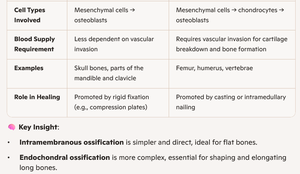
Blood Supply Requirement | Less dependent on vascular invasion | Requires vascular invasion for cartilage breakdown and bone formation |
Examples | Skull bones, parts of the mandible and clavicle | Femur, humerus, vertebrae |
Role in Healing | Promoted by rigid fixation (e.g., compression plates) | Promoted by casting or intramedullary nailing |
Key Insight: Intramembranous ossification is simpler and direct, ideal for flat bones. Endochondral ossification is more complex, essential for shaping and elongating long bones.
Longitudinal vs. Appositional Bone Growth
Bones grow in length and width through distinct processes.
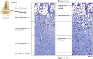
Longitudinal Growth: Occurs at the epiphyseal plates via chondrocyte proliferation, hypertrophy, calcification, and ossification. Responsible for increase in bone length during childhood and adolescence.
Appositional Growth: Increases bone width by adding new bone tissue beneath the periosteum; involves osteoblasts and osteoclasts for remodeling.
Hormonal Regulation of Bone Growth
Several hormones regulate bone growth and development.
Growth Hormone: Stimulates chondrocyte and osteoblast activity, increasing bone length and appositional growth.
Testosterone: Increases rate of longitudinal growth and accelerates closure of the epiphyseal plate.
Estrogen: Potently accelerates closure of the epiphyseal plate, ending longitudinal growth.
Bone Remodeling
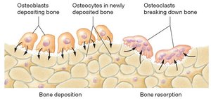
Bone remodeling is a continuous process of bone deposition and resorption, essential for:
Maintaining calcium ion (Ca2+) homeostasis
Bone repair
Replacing primary bone with secondary bone
Adapting bone to sustained tension and stresses
Osteoporosis
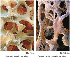
Osteoporosis is the most common bone disease in the United States, characterized by weakened, brittle bones due to inadequate inorganic matrix. It increases fracture risk and slows healing.
Causes: Dietary deficiencies (calcium, vitamin D), female gender, advanced age, lack of exercise, hormonal changes (e.g., postmenopausal estrogen loss), genetics, and other diseases.
Diagnosis: Bone density measurement.
Prevention: Balanced diet, supplementation, weight-bearing exercise, estrogen replacement if appropriate.
Treatment: Medications that inhibit osteoclasts or stimulate osteoblasts.