 Back
BackBones and Bone Structure: Study Notes for Anatomy & Physiology
Study Guide - Smart Notes
 Tailored notes based on your materials, expanded with key definitions, examples, and context.
Bones and Bone Structure
Functions of the Skeletal System
The skeletal system is a dynamic organ system that provides the framework for the human body. It consists of bones, cartilages, ligaments, and other connective tissues that stabilize and interconnect the bones.
Structural Support: The skeleton supports the body and maintains its shape.
Storage of Minerals and Lipids: Bones store minerals such as calcium and phosphate, as well as lipids in yellow bone marrow.
Blood Cell Production: Red bone marrow produces red blood cells, white blood cells, and platelets.
Protection: Bones protect soft tissues and vital organs (e.g., skull protects the brain, ribs protect the heart and lungs).
Leverage: Bones act as levers for muscles, enabling movement.
Classification of Bones
Bones are classified by their shapes and structures, each adapted for specific functions in the body.
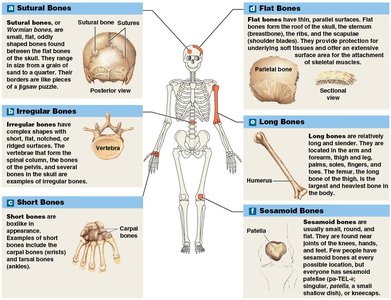
Sutural Bones (Wormian Bones): Small, flat, irregular bones found between the flat bones of the skull. Their number varies among individuals.
Irregular Bones: Complex shapes (e.g., vertebrae, pelvic bones).
Short Bones: Boxy in appearance (e.g., carpal and tarsal bones).
Flat Bones: Thin with parallel surfaces (e.g., skull roof, sternum, ribs, scapulae).
Long Bones: Long and slender (e.g., humerus, femur, bones of arms and legs).
Sesamoid Bones: Small, round, and flat bones found within tendons (e.g., patella). Their number and location can vary.
Bone Markings
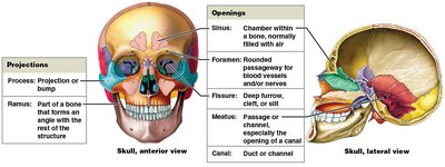
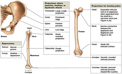
Bone markings are surface features that serve as sites for muscle, tendon, and ligament attachment, or as passages for nerves and blood vessels.
Projections: Sites for muscle and ligament attachment or articulation with other bones (e.g., process, ramus).
Openings and Depressions: Allow passage of blood vessels and nerves (e.g., foramen, canal, fissure, sinus, meatus, fossa, sulcus).
Structure of Bones
Long Bone Structure
Long bones have a characteristic structure that supports their function in movement and weight-bearing.
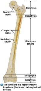
Diaphysis: The tubular shaft, composed of compact bone surrounding a medullary (marrow) cavity.
Epiphysis: The expanded ends, primarily made of spongy bone.
Metaphysis: The narrow region connecting diaphysis and epiphysis.
Flat Bone Structure
Flat bones consist of a core of spongy bone (diploë) sandwiched between two layers of compact bone (cortex).
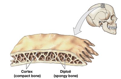
Bone Tissue (Osseous Tissue)
Bone tissue is a specialized connective tissue with a dense matrix of protein fibers and mineral deposits.
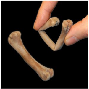
Matrix: Two-thirds is calcium phosphate (as hydroxyapatite crystals), providing hardness and resistance to compression. One-third is collagen fibers, providing flexibility and tensile strength.
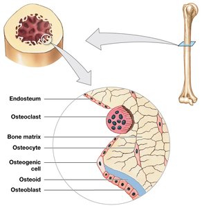
Osteocytes: Mature bone cells located in lacunae, connected by canaliculi for nutrient and waste exchange.
Periosteum: A membrane covering the outer surface of bones (except at joints), consisting of outer fibrous and inner cellular layers.
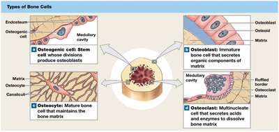
Types of Bone Cells
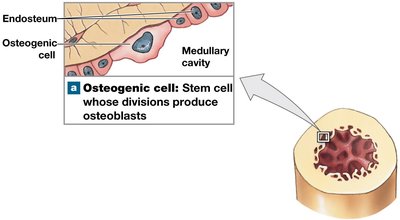
Osteogenic (Osteoprogenitor) Cells: Stem cells that divide to produce osteoblasts; important for fracture repair.
Osteoblasts: Immature bone cells responsible for osteogenesis (bone formation); secrete osteoid and promote mineralization.
Osteocytes: Mature bone cells that maintain the bone matrix and help repair damaged bone.
Osteoclasts: Large, multinucleate cells that resorb bone matrix (osteolysis) by secreting acids and enzymes.
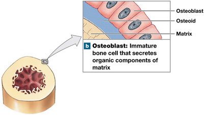
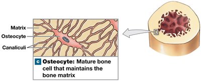
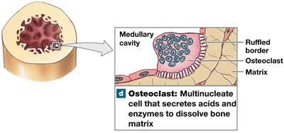
Compact Bone and Spongy Bone
Compact Bone
Compact bone is dense and forms the outer layer of bones, providing strength for weight-bearing.
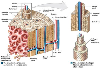
Osteon: The basic functional unit, consisting of concentric lamellae around a central canal containing blood vessels.
Lamellae: Layers of bone matrix; can be concentric (osteons), interstitial (between osteons), or circumferential (outer/inner surfaces).
Perforating (Volkmann's) Canals: Perpendicular to the surface, connecting blood vessels of osteons and periosteum.
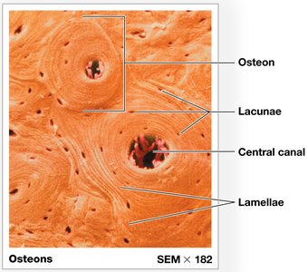
Spongy Bone
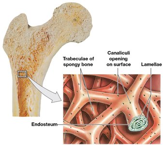
Spongy bone is composed of a network of trabeculae, making bones lighter and able to withstand stress from multiple directions. It supports and protects red bone marrow, which produces blood cells.
No osteons; matrix arranged as trabeculae.
Contains red bone marrow (hematopoiesis) and sometimes yellow bone marrow (fat storage).
Functional Integration
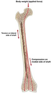
Compact and spongy bone work together to distribute forces and resist bending. For example, in the femur, the medial shaft compresses under weight, while the lateral shaft resists tension.
Surface Coverings
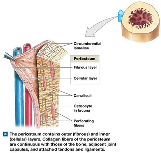
Periosteum: Outer membrane with fibrous and cellular layers; isolates bone, provides a route for blood vessels/nerves, and participates in growth and repair. Perforating (Sharpey's) fibers anchor it to bone.
Endosteum: Incomplete cellular layer lining inner surfaces (medullary cavity, trabeculae, central canals); active in bone growth, repair, and remodeling.
Bone Formation and Growth
Bone formation (ossification or osteogenesis) is the process of replacing other tissues with bone. Calcification is the deposition of calcium salts, necessary for ossification.
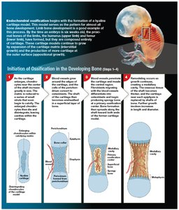
Endochondral Ossification: Bone replaces a cartilage model. Most bones form this way.
Intramembranous Ossification: Bone develops directly from mesenchymal tissue, forming dermal bones (e.g., flat bones of the skull).
Endochondral Ossification Steps
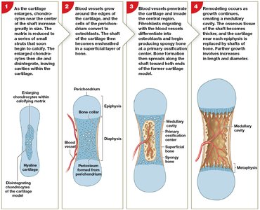
Chondrocytes enlarge, matrix calcifies, and chondrocytes die.
Blood vessels grow around cartilage; perichondrium cells become osteoblasts, forming a bone collar.
Blood vessels penetrate cartilage; fibroblasts become osteoblasts, forming spongy bone at the primary ossification center.
Remodeling creates a marrow cavity; bone thickens, cartilage at epiphyses replaced by bone.
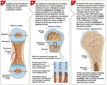
Secondary ossification centers form in epiphyses.
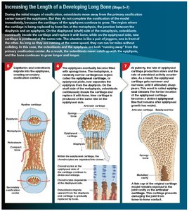
Epiphyses fill with spongy bone; epiphyseal plate separates diaphysis and epiphysis.
At puberty, epiphyseal closure occurs, leaving an epiphyseal line.
Bone Growth
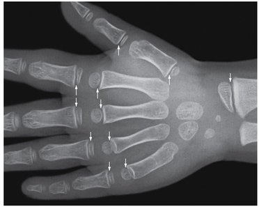
Interstitial Growth: Growth in length at the epiphyseal plate; ends with epiphyseal closure (epiphyseal line remains).
Appositional Growth: Growth in width; osteoblasts add layers to the outer surface, osteoclasts enlarge the medullary cavity.
Intramembranous Ossification Steps
Mesenchymal cells differentiate into osteoblasts, secrete osteoid, which calcifies.
Osteoblasts become osteocytes; bone grows in spicules.
Blood vessels grow between spicules; bone growth accelerates.
Spongy bone forms; outer layers remodel into compact bone; periosteum forms.
Bone Remodeling
Bone remodeling is the continuous recycling and renewal of bone matrix, involving osteocytes, osteoblasts, and osteoclasts. It maintains bone strength and mineral homeostasis.
Balanced activity keeps bones healthy; imbalance leads to weakened or strengthened bones.
Exercise, Nutrition, and Hormones
Bone health depends on physical activity, nutrition, and hormones.
Exercise: Stimulates bone remodeling and increases bone strength; inactivity leads to bone loss.
Nutrition: Requires adequate minerals (calcium, phosphorus, magnesium, fluoride, iron, manganese), vitamins (D, C, A, K, B12).
Hormones: Growth hormone, thyroxine, sex hormones, parathyroid hormone, and calcitonin regulate bone growth and calcium homeostasis.
Abnormal Bone Development: Includes pituitary growth failure, gigantism, acromegaly, and Marfan syndrome.
Calcium Homeostasis
Bones store 99% of the body's calcium, which is vital for physiological processes. Calcium levels are regulated by hormones:
Parathyroid Hormone (PTH): Increases blood calcium by stimulating osteoclasts, increasing intestinal absorption, and reducing kidney excretion.
Calcitonin: Decreases blood calcium by inhibiting osteoclasts, increasing kidney excretion, and reducing intestinal absorption.
Osteomalacia: Weak, flexible bones due to poor mineralization (e.g., rickets from vitamin D deficiency).
Fractures and Bone Repair
Fractures are cracks or breaks in bones due to physical stress. Types include open (compound), closed (simple), transverse, displaced, compression, spiral, epiphyseal, comminuted, greenstick, Colles, and Pott's fractures.
Repair Steps:
Fracture hematoma formation (blood clot seals area).
Callus formation (internal spongy bone and external cartilage/bone stabilize fracture).
Spongy bone formation (osteoblasts replace cartilage with spongy bone).
Compact bone formation (remodeling restores bone structure).
Effects of Aging on Bones
Aging leads to decreased bone mass and increased risk of fractures.
Osteopenia: Inadequate ossification, begins between ages 30-40; more pronounced in women.
Osteoporosis: Severe bone loss compromising function; common in postmenopausal women and older men; can be accelerated by hormonal changes or cancer.
