 Back
BackBones and Bone Tissue: Structure, Function, and Growth
Study Guide - Smart Notes
 Tailored notes based on your materials, expanded with key definitions, examples, and context.
Chapter 6: Bones & Bone Tissue
Functions of the Skeletal System
The skeletal system provides the framework for the body and serves several essential functions necessary for survival and movement.
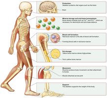
Protection: Bones protect vital organs, such as the brain (protected by the skull) and the heart and lungs (protected by the rib cage).
Mineral Storage and Acid-Base Homeostasis: Bones store minerals like calcium (Ca2+) and phosphate (PO43-), which are released into the blood as needed to maintain mineral balance and pH.
Blood Cell Formation: Hematopoiesis occurs in red bone marrow, producing red and white blood cells and platelets.
Fat Storage: Yellow bone marrow stores triglycerides, serving as an energy reserve.
Movement: Bones act as levers for muscles, enabling movement at joints.
Support: The skeleton supports the body’s weight and provides structural shape.
Components of the Skeletal System
Connective Tissues: Tendons, Ligaments, and Cartilage
The skeletal system is composed of bones and associated connective tissues, including tendons, ligaments, and cartilage.
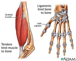
Tendons: Connect muscle to bone, transmitting the force needed for movement.
Ligaments: Connect bone to bone, stabilizing joints.
Cartilage: A resilient, semi-solid connective tissue found in joints, the ear, and other structures. Three types include:
Hyaline cartilage: Found at the ends of long bones, nose, trachea, and fetal skeleton.
Fibrocartilage: Found in intervertebral discs and menisci of the knee.
Elastic cartilage: Found in the ear and epiglottis.
Microscopic Structure of Tendons and Ligaments
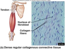
Tendons and ligaments are classified as dense regular collagenous connective tissue, which provides great tensile strength due to the abundance of collagen fibers.
Collagen: The primary protein, giving strength and flexibility.
Fibroblasts: The main cell type, responsible for producing collagen fibers.
Cartilage Structure
Cartilage is a specialized connective tissue with a semi-solid matrix. It contains:
Chondroblasts: Cells that produce the cartilage matrix.
Chondrocytes: Mature cartilage cells located in spaces called lacunae.
Matrix: Composed of collagen fibers (for strength) and proteoglycans (for resiliency).
Perichondrium: A double-layered connective tissue sheath covering cartilage (except at joints).
Comparison: Bone vs. Cartilage
Bones and cartilage differ in structure, function, and cellular composition.
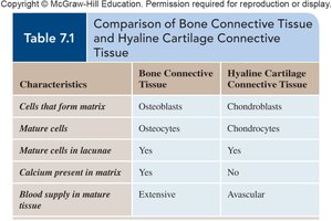
Characteristic | Bone Connective Tissue | Hyaline Cartilage Connective Tissue |
|---|---|---|
Cells that form matrix | Osteoblasts | Chondroblasts |
Mature cells | Osteocytes | Chondrocytes |
Mature cells in lacunae | Yes | Yes |
Calcium present in matrix | Yes | No |
Blood supply in mature tissue | Extensive | Avascular |
Classification of Bones
Types of Bones
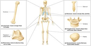
Bones are classified by shape, which relates to their function and location in the body.
Long bones: Longer than they are wide (e.g., humerus, femur, phalanges).
Short bones: About as long as they are wide (e.g., carpals, tarsals).
Flat bones: Thin, flat, and often curved (e.g., ribs, sternum, skull, scapulae).
Irregular bones: Complex shapes (e.g., vertebrae, facial bones).
Sesamoid bones: Small, round bones embedded in tendons (e.g., patella).
Structure of a Long Bone
Gross Anatomy of Long Bones
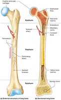
Long bones have a characteristic structure that supports their function in movement and weight-bearing.
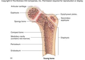
Diaphysis: The shaft, composed mainly of compact bone.
Epiphyses: The ends of the bone, containing spongy bone and covered with articular cartilage.
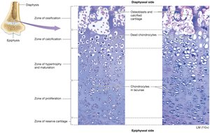
Epiphyseal Plate (Growth Plate): Hyaline cartilage region where bone growth occurs in children; becomes the epiphyseal line after growth stops.
Medullary Cavity: Central cavity containing red marrow (in children) or yellow marrow (in adults).
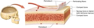
Periosteum: Membrane covering the outer surface, rich in blood vessels and nerves, anchored by perforating (Sharpey’s) fibers.
Compact vs. Spongy Bone
Long bones contain both compact and spongy bone, each with distinct properties.
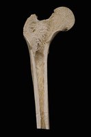
Compact bone: Dense outer layer that provides strength and resists compression and twisting.
Spongy bone (cancellous or trabecular): Internal network of bony struts, providing support and housing bone marrow.
Structure of Short, Flat, Irregular, and Sesamoid Bones
These bones lack a diaphysis and medullary cavity. They are covered by periosteum and consist of a sandwich of spongy bone between layers of compact bone. Some skull bones contain sinuses lined by mucous membranes.
Microscopic Structure of Bone Tissue
Bone Matrix Composition
The extracellular matrix (ECM) of bone is a composite material, providing both strength and flexibility.
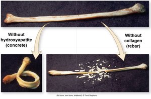
Organic component (35%): Collagen fibers and proteoglycans, providing tensile strength and flexibility.
Inorganic component (65%): Hydroxyapatite crystals (Ca10(PO4)6(OH)2), providing hardness and resistance to compression.
If hydroxyapatite is removed, bone becomes too flexible; if collagen is removed, bone becomes brittle.
Types of Bone Cells
Bone tissue contains several specialized cell types, each with distinct functions.
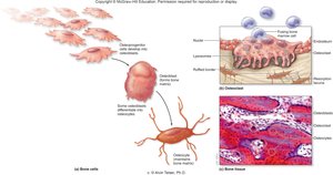
Osteogenic cells: Stem cells that differentiate into osteoblasts.
Osteoblasts: Bone-forming cells responsible for synthesizing bone matrix through ossification.
Osteocytes: Mature bone cells that maintain bone tissue; reside in lacunae and communicate via canaliculi.
Osteoclasts: Large, multinucleated cells responsible for bone resorption (breakdown), releasing minerals into the blood.
Microscopic Anatomy of Compact Bone
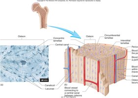
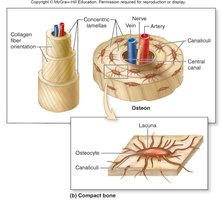
Compact bone is organized into osteons (Haversian systems), which are the basic functional units.
Osteon: Consists of a central canal surrounded by concentric lamellae (rings of bone matrix).
Central (Haversian) canal: Contains blood vessels and nerves.
Lacunae: Small spaces housing osteocytes.
Canaliculi: Tiny canals connecting lacunae, allowing nutrient and waste exchange.
Microscopic Anatomy of Spongy Bone
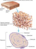
Spongy bone consists of a network of trabeculae ("little beams") oriented along lines of stress, with spaces filled by bone marrow and covered by endosteum.
Trabeculae: Lattice-like structures providing strength with minimal weight.
Marrow spaces: Contain red or yellow bone marrow.
Bone Formation and Growth
Ossification (Bone Formation)
Bone develops through two main processes: intramembranous and endochondral ossification.
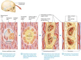
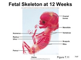
Intramembranous ossification: Forms flat bones of the skull, part of the mandible, and diaphyses of clavicles. Bone develops directly from mesenchymal tissue.
Endochondral ossification: Forms most bones below the head (except clavicles). Bone develops from a hyaline cartilage model.
Bone Growth in Length (Longitudinal/Interstitial Growth)
Long bones grow in length at the epiphyseal plate through the proliferation of cartilage, which is then replaced by bone.
Epiphyseal plate: Site of new cartilage formation and subsequent ossification.
When growth stops, the plate ossifies to become the epiphyseal line.
Bone Growth in Width (Appositional Growth)
Bones grow in width by the addition of new bone tissue at the surface, increasing the diameter of bones.
Osteoblasts: Add new bone to the periosteum.
Osteoclasts: Remove bone from the endosteum, enlarging the medullary cavity.
Bone Remodeling and Homeostasis
Bone Remodeling
Bone is continuously remodeled throughout life, balancing bone formation and resorption to adapt to stress and maintain mineral homeostasis.
Osteoblasts: Build new bone matrix.
Osteoclasts: Break down bone matrix, releasing minerals.
Remodeling is influenced by mechanical stress, hormones, and nutritional status.
Calcium Homeostasis
Bone acts as a reservoir for calcium, which is critical for nerve and muscle function, blood clotting, and enzyme activity. Blood calcium levels are regulated by hormones:
Parathyroid hormone (PTH): Increases blood calcium by stimulating osteoclast activity, increasing intestinal absorption, and reducing renal excretion.
Calcitonin (CT): Lowers blood calcium by inhibiting osteoclasts and increasing calcium deposition in bone.
Bone Fractures and Repair
Types of Bone Fractures
Bone fractures are classified by their characteristics and causes:
Stress fracture: Caused by repetitive force or overuse.
Pathologic fracture: Occurs in bone weakened by disease.
Simple (closed) fracture: Bone is broken but does not penetrate the skin.
Compound (open) fracture: Bone pierces the skin, increasing risk of infection.
Fracture Repair
Bone repair occurs in four main steps:
Bleeding and formation of a fracture hematoma (clot).
Formation of a fibrocartilaginous (soft) callus.
Formation of a bony (hard) callus.
Bone remodeling restores the bone’s original shape and structure.
Bone Disorders and Aging
Osteopenia and Osteoporosis
Bone mass decreases with age, leading to osteopenia (mild bone loss) and osteoporosis (severe bone loss), which increase fracture risk. Osteoporosis is more common in postmenopausal women due to decreased estrogen.
Prevention and treatment: Weight-bearing exercise, adequate calcium and vitamin D intake, and medications can help maintain bone density.
Effects of Aging on the Skeletal System
With aging, bone matrix decreases, bones become more brittle, and bone mass declines. This leads to increased risk of fractures, deformity, loss of height, pain, and stiffness.
