 Back
BackBones and Bone Tissue: Structure, Function, and Cellular Anatomy
Study Guide - Smart Notes
 Tailored notes based on your materials, expanded with key definitions, examples, and context.
Ch. 6 Bones and Bone Tissue
Introduction to Bone and Skeletal Tissue
The skeletal system is composed of bones and associated tissues, forming a dynamic organ system that responds to its environment. Bones serve multiple essential functions in the human body.
Support: Bones provide structural support for the body, maintaining its shape and posture.
Protection: Bones protect soft internal organs, such as the brain (cranium), heart and lungs (rib cage), and spinal cord (vertebrae).
Production of Blood Cells: Bones are the site of hematopoiesis, the formation of blood cells.
Storage: Bones store fat (triglycerides) and minerals, primarily calcium and phosphate.
Levers for Muscles: Bones act as levers, enabling movement when muscles contract.
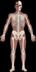


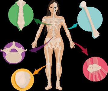
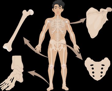
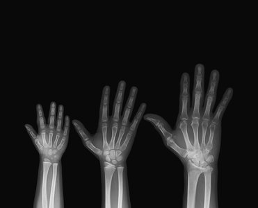
Types of Bones
Bones are classified based on their shape and structure, which relates to their function and location in the body.
Long Bones: Shaped like a rod, with expanded ends. Examples: humerus, femur, phalanges.
Short Bones: Cube-shaped. Examples: wrist (carpals), ankle (tarsals).
Flat Bones: Thin, flat, and slightly curved. Examples: sternum, ribs, cranial bones.
Irregular Bones: Complex shapes. Examples: vertebrae, pelvis, facial bones.
Sesamoid Bones: Develop within a tendon, variable in number. Example: patella.
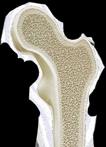
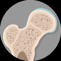
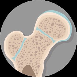
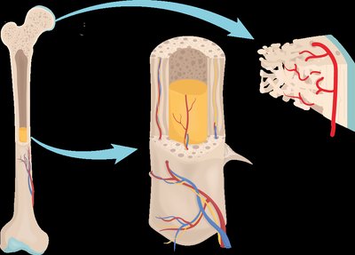
Structural Arrangements: Compact and Spongy Bone
All bones have two main structural arrangements: compact bone and spongy bone. These arrangements provide strength and flexibility.
Compact Bone: Appears solid, with no visible spaces. Optimized for strength and hardness. Found at the edges of all bones and in the shaft of long bones.
Spongy Bone: Appears porous, built like scaffolding. Contains trabeculae (bone struts), reduces weight, and spaces are filled with marrow. Found in the interior of bones.

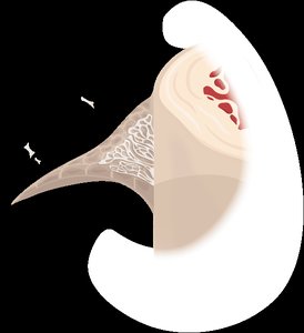
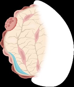
Gross Anatomy of Bone: Periosteum and Endosteum
Bones are covered and lined by specialized connective tissues that provide nourishment, attachment points, and house bone stem cells.
Periosteum: Covers the external surface of bone; consists of two layers (fibrous and osteogenic). Highly vascularized and innervated. Collagen fibers from tendons and ligaments weave into the fibrous layer, forming strong connections.
Endosteum: Lines all internal surfaces of bone; similar composition to the osteogenic layer of the periosteum.
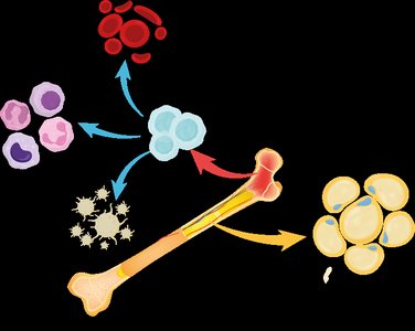
Gross Anatomy of Bone: Bone Marrow
The spaces inside bones are filled with bone marrow, which exists in two main types: red and yellow marrow.
Red Marrow: Site of hematopoiesis (formation of blood cells). Found in spongy bone in adults; primary marrow type in infants.
Yellow Marrow: Stores fat (triglycerides). Found in spongy bone and medullary cavity; primary marrow type in adults. Can revert to red marrow if needed.


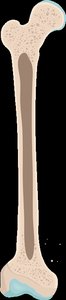
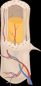
Gross Anatomy of Bones: Structure of a Long Bone
Long bones have a distinct structure, consisting of two ends (epiphyses) and a shaft (diaphysis).
Epiphysis: Wider end of a long bone, covered with articular cartilage at joints.
Diaphysis: Tubular shaft of the bone, contains the medullary cavity filled with yellow marrow.
Metaphysis: Area where epiphysis and diaphysis meet; contains the epiphyseal plate (site of bone growth until adulthood).
Epiphyseal Line: Plate converts to bone when growth is complete.
Nerves and Blood Supply
Bones contain blood vessels and nerves, which enter through small openings called nutrient foramina.
Nutrient Foramen: Small hole in diaphysis for blood vessels and nerves.
Nutrient Artery: Brings blood into the medullary cavity.
Nutrient Vein: Carries blood out of the bone.
Smaller Foramina: Located in metaphysis and epiphysis.
Microscopic Anatomy of Bones: Bone Matrix
The extracellular matrix of bone consists of two basic components: inorganic and organic matrices.
Inorganic Matrix: Hydroxyapatite crystals (calcium and phosphate), making up about 65% of bone mass. Provides hardness and rigidity.
Organic Matrix (Osteoid): Collagen fibers and ground substance, making up about 35% of bone mass. Provides strength and flexibility.
Equation:
Microscopic Anatomy of Bones: Bone Cells
Bone tissue is comprised of several cell types, each with distinct functions in bone formation, maintenance, and remodeling.
Osteoprogenitor (Osteogenic) Cells: Bone stem cells found in periosteum and endosteum.
Osteoblasts: Build bone by secreting bone matrix and collagen fibers. Arise from osteoprogenitor cells.
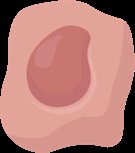
Osteocytes: Mature bone cells that maintain the matrix. Trapped in lacunae, monitor bone stress, and contribute to calcium homeostasis.
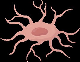
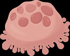
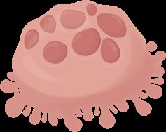
Osteoclasts: Break down bone for remodeling. Multinucleate, derived from white blood cells, have a ruffled border to increase surface area for bone resorption.


Microscopic Anatomy of Bones: The Osteon
The osteon (Haversian system) is the structural unit of compact bone, consisting of concentric lamellae, central canals, and canaliculi.
Central Canal: Contains blood vessels and nerves, runs parallel to the axis of the bone.
Perforating Canals: Run perpendicular to central canals, connecting them to other blood vessels and nerves.
Canaliculi: Small channels for communication and transport between osteocytes.
Lacunae: Chambers containing osteocytes.
Lamellae: Concentric rings of matrix.
Microscopic Anatomy of Bones: Lamellae and Collagen Arrangement
Collagen fibers within lamellae are arranged in alternating directions, providing strength and resistance to stress from multiple directions.
Within Lamella: Collagen runs in one direction.
Between Adjacent Lamellae: Collagen runs in alternate directions.
Effect: Provides strength and resists twisting forces.
Microscopic Anatomy of Bones: Trabeculae
Spongy bone is characterized by a lattice-like appearance with many open spaces. Trabeculae are small rods or struts in spongy bone, containing lamellae, osteocytes, and canaliculi, but lacking well-organized osteons and central canals.
Trabeculae: Align with lines of stress, providing structural support.
Spongy Bone: Also called cancellous bone.
Summary Table: Compact vs. Spongy Bone Structures
Structure | Compact Bone | Spongy Bone |
|---|---|---|
Osteons | Present | Absent |
Trabeculae | Absent | Present |
Central Canals | Present | Absent |
Perforating Canals | Present | Absent |
Endosteum | Present | Present |
Osteocytes | Present | Present |
Canaliculi | Present | Present |
Additional info: Academic context was added to clarify bone cell functions, structural arrangements, and the relationship between bone matrix components and bone strength.
