 Back
Ch. 6 - Bones and Bone Tissue: Structure, Function, and Physiology
Study Guide - Smart Notes
 Tailored notes based on your materials, expanded with key definitions, examples, and context.
Bones and Bone Tissue
Overview of the Skeletal System
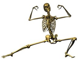
The skeletal system is a complex organ system composed of bones, joints, and associated supporting structures such as cartilage, ligaments, and tendons. Bones are the primary organs of this system and are made of two types of osseous tissue: spongy bone and compact bone. In addition, bones contain other connective tissues, including membranes and bone marrow.
Functions of the Skeletal System
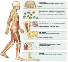
Protection: Bones protect vital organs such as the brain, heart, and lungs.
Mineral Storage and Acid-Base Homeostasis: Bones store minerals such as calcium (Ca2+) and phosphate (PO43-), which are essential for electrolyte and acid-base balance.
Blood Cell Formation: Red bone marrow is the site of hematopoiesis, the formation of blood cells.
Fat Storage: Yellow bone marrow stores triglycerides, serving as an energy reserve.
Movement: Muscles produce movement by pulling on bones.
Support: The skeleton supports the weight of the body and provides its structural framework.
Cartilage, Tendons, and Ligaments
Cartilage Structure and Function
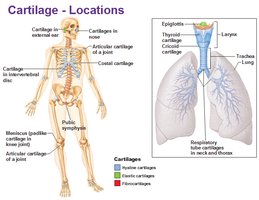
Cartilage is a specialized connective tissue consisting of cells that produce a matrix. The main cell types are chondroblasts (which form the matrix) and chondrocytes (which are surrounded by the matrix in spaces called lacunae). The matrix is rich in ground substance, collagen for strength, and elastin for resiliency. Most cartilage is covered by a double-layered sheath called the perichondrium, except at articulations (joints).
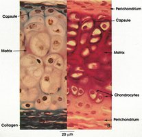
Articular cartilage: Covers bones at joints and lacks a perichondrium.
Growth types:
Appositional growth: New chondrocytes and matrix are added at the periphery.
Interstitial growth: Chondrocytes within the tissue divide and add more matrix between cells.
Tendons and Ligaments
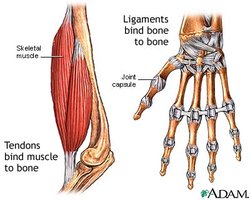
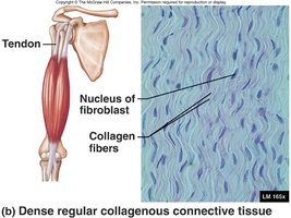
Tendons and ligaments are types of dense regular collagenous connective tissue. Tendons connect muscle to bone, while ligaments connect bone to bone. Their strength is due to the high content of collagen fibers.
Classification and Structure of Bones
Bone Classification by Shape
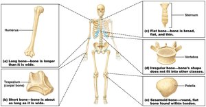
Humans have approximately 206 bones, which are classified into five categories based on shape:
Long bones: Longer than they are wide (e.g., humerus).
Short bones: About as long as they are wide (e.g., carpals).
Flat bones: Broad, flat, and thin (e.g., sternum).
Irregular bones: Complex shapes (e.g., vertebrae).
Sesamoid bones: Round, flat bones found within tendons (e.g., patella).
Bone Tissues: Compact and Spongy Bone
There are two main types of bone tissue:
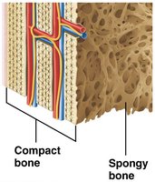
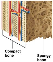
Compact bone: Hard, dense outer region that resists linear compression and twisting forces.
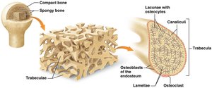
Spongy bone (cancellous bone): Porous framework of trabeculae found deep in bone, allowing resistance to forces from many directions and providing cavities for bone marrow.
Structure of Long Bones
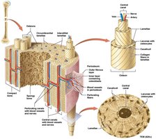
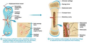
Long bones have a central shaft called the diaphysis (mostly compact bone) and expanded ends called epiphyses (mostly spongy bone). The diaphysis contains the medullary cavity filled with yellow bone marrow. The epiphyses are covered with articular cartilage and surrounded by the periosteum, a membrane rich in blood vessels and nerves, attached by perforating fibers (Sharpey's fibers). The endosteum lines the internal surfaces and contains bone cells involved in homeostasis. The epiphyseal line is a remnant of the growth plate.
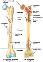
Structure of Short, Flat, Irregular, and Sesamoid Bones
These bones lack diaphyses, epiphyses, medullary cavities, and epiphyseal lines/plates. They are covered by periosteum and have two outer layers of compact bone with a middle layer of spongy bone called diploë. Some flat and irregular bones contain sinuses to reduce weight.
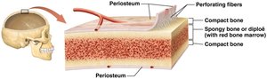
Blood and Nerve Supply to Bone
Blood supply to bones is provided by vessels in the periosteum and nutrient arteries that enter through nutrient foramina. Short, flat, irregular, and sesamoid bones are mainly supplied by periosteal vessels, while long bones receive additional supply from nutrient arteries.
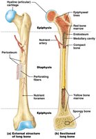
Bone Marrow
Red bone marrow contains hematopoietic stem cells and is the site of blood cell formation. In adults, it is found in the pelvis, proximal femur and humerus, vertebrae, ribs, sternum, clavicles, scapulae, and some skull bones. Yellow bone marrow consists of adipocytes and blood vessels and serves as an energy reserve. Red marrow is used in bone marrow transplants for conditions like leukemia.
Microscopic Structure of Bone
Bone Matrix
The extracellular matrix (ECM) of bone consists of:
Inorganic matrix (65%): Mainly calcium salts, especially hydroxyapatite crystals (), providing strength and resistance to compression.
Organic matrix (35%): Also called osteoid, composed of collagen fibers, proteoglycans, glycosaminoglycans, glycoproteins, and bone-specific proteins. Collagen provides tensile strength, while proteoglycans and glycosaminoglycans help resist compression by drawing water into the matrix.
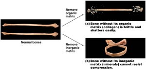
Bone Cells
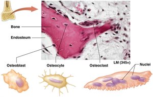
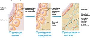
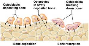
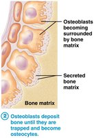
Osteoblasts: Bone-building cells found in periosteum and endosteum; secrete bone matrix.
Osteocytes: Mature bone cells that maintain bone matrix; reside in lacunae.
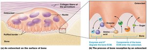
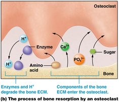
Osteoclasts: Large, multinucleated cells that break down bone matrix (resorption); derived from bone marrow stem cells.
Histology of Compact and Spongy Bone
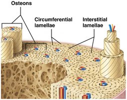
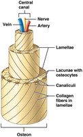
Compact bone is organized into osteons (Haversian systems), which are cylindrical structures composed of concentric lamellae. Each osteon has a central canal containing blood vessels and nerves. Osteocytes reside in lacunae between lamellae and communicate via canaliculi. Perforating (Volkmann's) canals connect osteons to the periosteum and other osteons. Spongy bone consists of trabeculae, which are oriented to resist stress from multiple directions and house bone marrow.
Bone Development and Growth
Ossification (Osteogenesis)
Bone formation begins in the embryo and continues through childhood. There are two mechanisms:
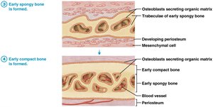
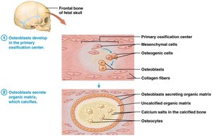
Intramembranous ossification: Forms flat bones (e.g., skull, clavicles) from a membrane of embryonic mesenchymal tissue.
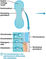
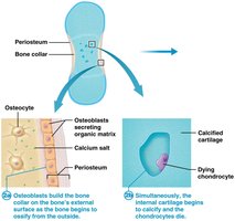
Endochondral ossification: Forms most bones below the head (except clavicle) from a hyaline cartilage model.
Growth in Length (Longitudinal Growth)
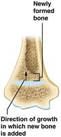
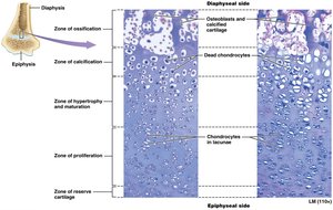
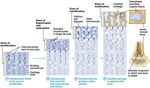
Longitudinal growth occurs at the epiphyseal plate through the division of chondrocytes. The plate consists of several zones:
Zone of reserve cartilage: Inactive chondrocytes closest to the epiphysis.
Zone of proliferation: Actively dividing chondrocytes.
Zone of hypertrophy and maturation: Enlarged, non-dividing chondrocytes.
Zone of calcification: Dead chondrocytes and calcified matrix.
Zone of ossification: Osteoblasts replace calcified cartilage with bone.
Growth in Width (Appositional Growth)
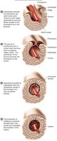
Appositional growth increases bone width as osteoblasts in the periosteum lay down new bone, forming circumferential lamellae. Bones can continue to grow in width even after longitudinal growth has ceased.

Hormonal Regulation of Bone Growth
Hormones such as growth hormone (GH), testosterone, and estrogen regulate bone growth. GH stimulates chondrocyte division and osteoblast activity, promoting both longitudinal and appositional growth. Testosterone increases bone thickness and calcium deposition, while estrogen accelerates epiphyseal plate closure, resulting in shorter stature in females.

Bone Growth Pathophysiologies
Gigantism: Excess GH in childhood leads to excessive bone growth.
Acromegaly: Excess GH in adulthood causes thickening of bones and soft tissues.
Achondroplasia: Genetic disorder affecting endochondral ossification, resulting in short limbs and abnormal bone growth.
Bone Remodeling and Homeostasis
Bone Remodeling
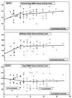
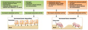
Bone remodeling is a continuous process involving bone deposition by osteoblasts and resorption by osteoclasts. Functions include calcium homeostasis, bone repair, adaptation to stress, and replacement of old bone.
Calcium Homeostasis
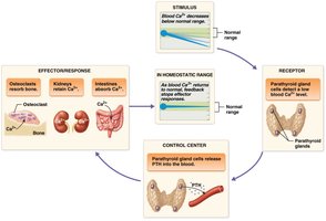
Calcium levels in the blood are regulated by a negative feedback loop involving the thyroid and parathyroid glands. Parathyroid hormone (PTH) increases blood calcium by stimulating osteoclast activity, reducing urinary calcium loss, and activating vitamin D. Calcitonin from the thyroid decreases blood calcium by stimulating osteoblast activity and increasing urinary calcium loss.
Bone Injuries and Repair
Bone Fractures
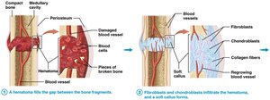
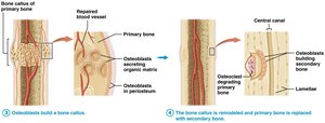
Fractures are breaks in bone. Simple fractures do not break the skin, while compound fractures do. Healing involves four steps: hematoma formation, soft callus formation, hard callus formation, and bone remodeling.
Pathophysiology and Aging of the Skeletal System
Osteoporosis
Osteoporosis is a common bone disease characterized by weak and brittle bones due to inadequate inorganic matrix. Risk factors include poor diet, female gender, advanced age, lack of exercise, hormonal changes, and genetics. Prevention and treatment involve diet, exercise, and medications that affect osteoclast and osteoblast activity.
Aging and the Skeletal System
With age, bone matrix decreases, leading to increased brittleness and decreased bone mass. Bone loss accelerates after menopause, especially in spongy bone, increasing the risk of fractures, deformity, and loss of height.
