 Back
BackBones and Bone Tissue: Structure, Function, and Growth
Study Guide - Smart Notes
 Tailored notes based on your materials, expanded with key definitions, examples, and context.
Bones and Bone Tissue
Introduction to Bones as Organs
The skeletal system is composed of bones, joints, and associated supporting tissues. Bones are the main organs of the system and are made up of more than just osseous tissue; they also contain dense regular and irregular collagenous connective tissue and bone marrow.
Functions of the Skeletal System
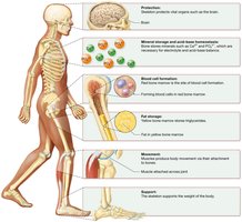
Protection: Bones such as the skull, sternum, ribs, and pelvis protect underlying organs, exemplifying the structure-function core principle.
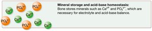
Mineral Storage and Acid–Base Homeostasis: Bones are the primary storehouse for calcium, phosphorus, and magnesium salts, which are critical for electrolyte and acid–base balance.
Blood Cell Formation: Red bone marrow is the site of hematopoiesis, the formation of blood cells.
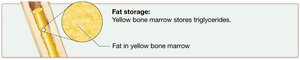
Fat Storage: Yellow bone marrow stores triglycerides, which can be used as an energy source.
Movement: Bones serve as attachment sites for skeletal muscles; muscle contraction pulls on bones to generate movement at joints.
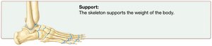
Support: The skeleton supports the body’s weight and provides its structural framework.



Classification of Bones by Shape
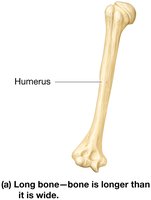
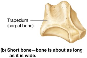
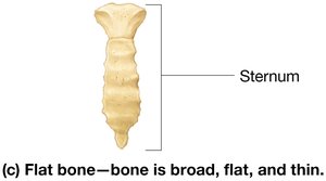
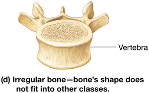
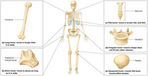
Bones are classified into five categories based on their shape, not size:
Long Bones: Longer than they are wide (e.g., humerus, femur).
Short Bones: Roughly cube-shaped, as long as they are wide (e.g., carpals, tarsals).
Flat Bones: Thin, broad, and usually curved (e.g., sternum, ribs, skull bones).
Irregular Bones: Complex shapes that do not fit other categories (e.g., vertebrae, certain skull bones).
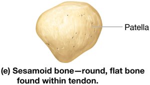
Sesamoid Bones: Small, flat, oval-shaped bones within tendons (e.g., patella).
Structure of Long Bones
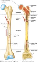
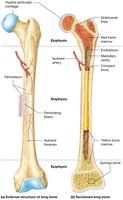
Long bones have a unique structure that supports their function:
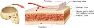
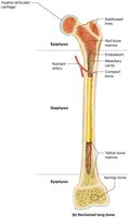
Periosteum: Dense irregular collagenous connective tissue membrane covering the outer surface, rich in blood vessels and nerves.
Perforating (Sharpey’s) Fibers: Collagen fibers anchoring periosteum to bone.
Diaphysis: Shaft of the bone.
Epiphyses: Ends of the bone, covered with articular (hyaline) cartilage.
Medullary Cavity: Central cavity containing red or yellow bone marrow.
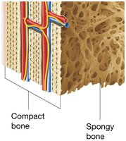
Compact Bone: Dense outer region providing strength and resistance to compression and twisting.
Spongy Bone (Cancellous Bone): Inner honeycomb-like structure that resists forces from multiple directions and houses bone marrow.
Endosteum: Thin membrane lining internal surfaces, involved in bone homeostasis.
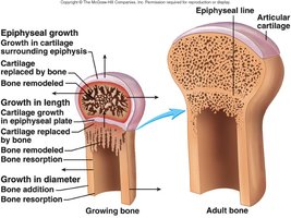
Epiphyseal Line: Remnant of the epiphyseal plate (growth plate), marking where bone growth occurred in children.
Structure of Other Bone Types
Short, flat, irregular, and sesamoid bones do not have diaphyses, epiphyses, or medullary cavities. They are covered by periosteum and consist of two layers of compact bone with a middle layer of spongy bone (diploë). Some skull bones contain sinuses to reduce weight.
Blood and Nerve Supply to Bone
Short, flat, irregular, and sesamoid bones receive blood from periosteal vessels.
Long bones receive one-third of their blood from periosteum and two-thirds from nutrient arteries entering through the nutrient foramen.
Epiphyses are supplied by small blood vessels entering through compact bone.
Bone Marrow
Red Bone Marrow: Contains reticular fibers and hematopoietic cells; site of blood cell formation. More abundant in children, restricted to certain bones in adults (pelvis, proximal femur/humerus, vertebrae, ribs, sternum, clavicles, scapulae, some skull bones).
Yellow Bone Marrow: Stores triglycerides, mainly composed of adipocytes and blood vessels. Replaces red marrow with age but can revert to red marrow if needed (e.g., severe blood loss).
Microscopic Structure of Bone Tissue
Bone (osseous tissue) is primarily extracellular matrix with a small population of cells. The matrix is divided into:
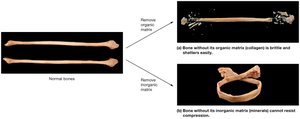
Inorganic Matrix: About 65% of bone weight, mainly hydroxyapatite crystals (calcium and phosphate), providing hardness and resistance to compression.
Organic Matrix (Osteoid): About 35% of bone weight, mainly collagen fibers, proteoglycans, glycosaminoglycans, glycoproteins, and bone-specific proteins, providing flexibility and tensile strength.
Bone Cells
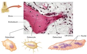
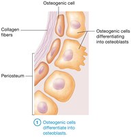
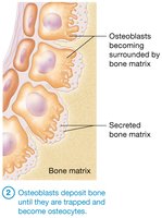
Osteoblasts: Bone-building cells found in periosteum and endosteum; secrete organic matrix and assist in inorganic matrix formation (bone deposition).
Osteocytes: Mature bone cells trapped in lacunae; maintain bone matrix and can recruit osteoblasts for repair.
Osteoclasts: Large, multinucleated cells responsible for bone resorption; secrete hydrogen ions and enzymes to break down bone matrix.
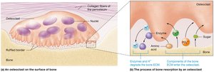
Histology of Bone
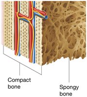
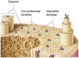
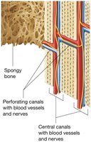
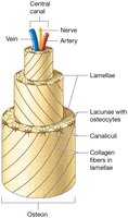
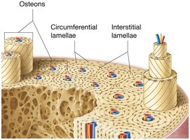
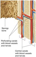
Compact Bone: Composed of osteons (Haversian systems) with concentric lamellae, central canals, and canaliculi connecting osteocytes. Interstitial and circumferential lamellae add strength.
Spongy Bone: Composed of trabeculae with concentric lamellae and osteocytes; lacks central canals, receives nutrients from bone marrow vessels.
Bone Formation: Ossification
Ossification (osteogenesis) is the process of bone formation, beginning in the embryonic period and continuing through childhood. Two mechanisms exist:
Intramembranous Ossification: Forms flat bones (e.g., skull, clavicles) from a membrane of embryonic connective tissue (mesenchymal cells).
Endochondral Ossification: Forms most bones below the head (except clavicles) from a hyaline cartilage model.
Intramembranous Ossification Steps
Mesenchymal cells differentiate into osteogenic cells, then osteoblasts at the primary ossification center.
Osteoblasts secrete organic matrix; calcification occurs as calcium salts are deposited.
Osteoblasts trapped in matrix become osteocytes.
Early spongy bone forms, then periosteum develops, and compact bone forms on the outer layers.
Endochondral Ossification Steps
Chondroblasts in perichondrium differentiate into osteogenic cells, then osteoblasts, forming periosteum.
Bone collar forms on external surface; internal cartilage calcifies and chondrocytes die.
Osteoclasts create openings for blood vessels; osteoblasts replace calcified cartilage with spongy bone.
Secondary ossification centers develop in epiphyses; cartilage remains at epiphyseal plates and articular surfaces.
Bone Growth in Length and Width
Longitudinal Growth: Occurs at the epiphyseal plate via chondrocyte division and ossification. The plate has five zones: reserve cartilage, proliferation, hypertrophy/maturation, calcification, and ossification.
Appositional Growth: Bones grow in width as osteoblasts lay down new bone at the periosteum, forming new circumferential lamellae.
Bone Remodeling and Repair
Bone Remodeling: Continuous process involving bone deposition (by osteoblasts) and resorption (by osteoclasts). Maintains calcium homeostasis, adapts to stress, and replaces old bone.
Calcium Homeostasis: Regulated by parathyroid hormone (PTH) and calcitonin in a negative feedback loop. PTH increases blood calcium by stimulating osteoclasts, increasing gut absorption, and reducing urinary loss. Calcitonin promotes bone deposition.
Bone Repair: After a fracture, a hematoma forms, followed by a soft callus (fibrocartilage), then a hard callus (primary bone), which is remodeled into secondary bone.
Table: Types of Bone Cells
Cell Type | Location | Function |
|---|---|---|
Osteoblast | Periosteum, endosteum | Bone deposition |
Osteocyte | Lacunae in bone matrix | Maintain bone matrix |
Osteoclast | Bone surfaces | Bone resorption |
Table: Classification of Bones by Shape
Type | Shape | Example |
|---|---|---|
Long | Longer than wide | Humerus, femur |
Short | Cube-shaped | Carpals, tarsals |
Flat | Thin, broad | Sternum, ribs |
Irregular | Complex shape | Vertebrae |
Sesamoid | Small, oval, within tendons | Patella |
Additional info: This guide covers the essential structure, function, and growth of bone tissue, as well as the cellular and molecular mechanisms underlying bone physiology. It is suitable for exam preparation in a college-level Anatomy & Physiology course.
