 Back
BackBones and Skeletal Tissue: Structure, Function, and Classification
Study Guide - Smart Notes
 Tailored notes based on your materials, expanded with key definitions, examples, and context.
Bones and Skeletal Tissue
Types of Skeletal Cartilage
The human skeleton is initially composed of cartilage, which is gradually replaced by bone during development. Cartilage is a resilient connective tissue that provides support and flexibility in various parts of the body. There are three primary types of cartilage:
Hyaline cartilage: Most abundant; provides support, flexibility, and resilience. Found in articular surfaces of joints, costal cartilage (ribs), respiratory structures (larynx), and nasal cavities.
Elastic cartilage: Similar to hyaline but contains elastic fibers, allowing greater flexibility. Located in the external ear and epiglottis.
Fibrocartilage: Contains thick collagen fibers for tensile strength. Found in menisci of the knee, intervertebral discs, and pubic symphysis.
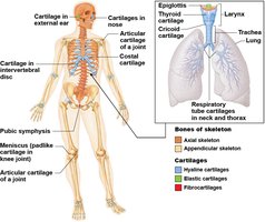
Structure and Properties of Skeletal Cartilage
Skeletal cartilage is highly resilient due to its high water content and lacks blood vessels and nerves. The main cell type is the chondrocyte, which resides in spaces called lacunae. Cartilage is surrounded by the perichondrium, a dense connective tissue layer that resists outward expansion and contains blood vessels for nutrient delivery.
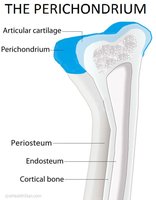
Growth of Cartilage
Cartilage grows by two mechanisms:
Appositional growth: Cartilage-forming cells in the perichondrium secrete new matrix on the external surface of existing cartilage.
Interstitial growth: Chondrocytes within lacunae divide and secrete new matrix, expanding cartilage from within.
Functions of Bone
Major Functions
Bones serve multiple essential functions in the human body:
Support: Provide structural framework for the body and soft organs.
Protection: Shield vital organs such as the brain, spinal cord, and thoracic organs.
Movement: Act as levers for muscle action.
Mineral and growth factor storage: Store calcium, phosphorus, and growth factors.
Blood cell formation: Hematopoiesis occurs in red marrow cavities.
Triglyceride storage: Fat stored in bone cavities serves as an energy reserve.
Hormone production: Osteocalcin regulates insulin secretion, glucose levels, and metabolism.
Classification of Bones
Axial and Appendicular Skeleton
The 206 named bones in the human skeleton are divided into two groups:
Axial skeleton: Long axis of the body; includes skull, vertebral column, and rib cage.
Appendicular skeleton: Bones of the limbs and girdles attaching limbs to the axial skeleton.
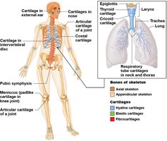
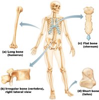
Bone Shapes
Bones are classified by shape:
Long bones: Longer than wide; found in limbs.
Short bones: Cube-shaped; found in wrist and ankle. Sesamoid bones form within tendons.
Flat bones: Thin, flat, and slightly curved; includes sternum, scapulae, ribs, and most skull bones.
Irregular bones: Complex shapes; includes vertebrae and hip bones.
Bone Structure
Levels of Bone Structure
Bones are organs composed of bone (osseous) tissue, nervous tissue, connective tissue, and epithelium. Structure is studied at three levels:
Gross anatomy
Microscopic anatomy
Chemical composition
Gross Anatomy: Compact and Spongy Bone
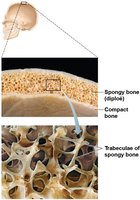
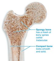
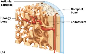
All bones have a dense outer layer called compact bone and an internal honeycomb structure called spongy bone, which consists of trabeculae. The spaces between trabeculae are filled with red or yellow bone marrow.
Structure of Short, Irregular, and Flat Bones
These bones consist of thin plates of spongy bone (diploë) covered by compact bone. The periosteum covers the outside, and the endosteum covers the inside. Bone marrow is scattered throughout the spongy bone, and hyaline cartilage covers areas involved in joints.
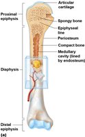
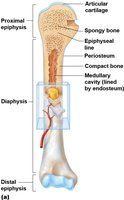
Structure of Typical Long Bone
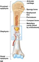
Long bones have three main regions:
Diaphysis: Tubular shaft forming the long axis, composed of compact bone surrounding the medullary cavity (filled with yellow marrow in adults).
Epiphyses: Ends of the bone, consisting of compact bone externally and spongy bone internally. Articular cartilage covers joint surfaces.
Metaphysis: Flared region where diaphysis and epiphysis meet; contains the epiphyseal line (remnant of the growth plate).
Bone Membranes
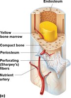
Bones are covered by two membranes:
Periosteum: Double-layered membrane covering external surfaces except joints. The fibrous layer consists of dense irregular connective tissue and Sharpey's fibers; the osteogenic layer contains stem cells for bone growth and repair. It is richly supplied with nerves and blood vessels.
Endosteum: Delicate membrane covering internal bone surfaces and trabeculae of spongy bone; also contains osteogenic stem cells.
Hematopoietic Tissue in Bone
Red marrow is found in trabecular cavities of spongy bone and diploë of flat bones. In adults, it is located in the heads of femur and humerus, as well as flat and some irregular bones. Yellow marrow can convert to red marrow if needed (e.g., in anemia).
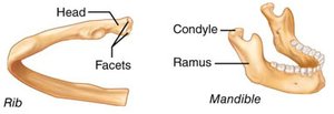
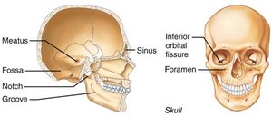
Bone Markings
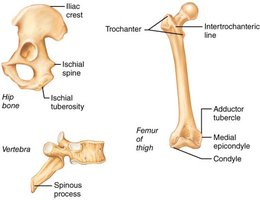
Bones have markings that serve as sites for muscle, ligament, and tendon attachment, or as passageways for blood vessels and nerves. Markings are classified as:
Projection: Outward bulge of bone.
Depression: Bowl- or groove-like cut out.
Opening: Hole or canal serving as passageways.
Summary Table: Types of Cartilage and Their Locations
Type of Cartilage | Main Features | Locations |
|---|---|---|
Hyaline | Support, flexibility, resilience | Joints, ribs, respiratory tract, nose |
Elastic | Elastic fibers, flexibility | External ear, epiglottis |
Fibrocartilage | Thick collagen, tensile strength | Knee menisci, intervertebral discs, pubic symphysis |
Summary Table: Bone Classification
Bone Shape | Examples |
|---|---|
Long | Femur, humerus |
Short | Carpals, tarsals |
Flat | Sternum, scapulae, ribs, skull |
Irregular | Vertebrae, hip bones |
Key Equations
Bone growth and remodeling are regulated by hormones and mechanical stress. The following equation represents the relationship between bone mass and mineral deposition:
Additional info: Bone remodeling is a continuous process involving osteoblasts (bone-forming cells) and osteoclasts (bone-resorbing cells).
