 Back
BackBones and Skeletal Tissues: Structure, Function, and Development
Study Guide - Smart Notes
 Tailored notes based on your materials, expanded with key definitions, examples, and context.
Bones and Skeletal Tissues
Introduction to the Skeletal System
The skeletal system forms the structural framework of the human body, providing support, protection, and facilitating movement. It is composed of bones and associated skeletal cartilages, which undergo development and remodeling throughout life.
Functions of the Skeletal System
Major Functions of Bones
Support: Bones provide the rigid framework that supports the body and cradles soft organs.
Protection: The skeleton protects vital organs, such as the brain (skull), spinal cord (vertebrae), and thoracic organs (rib cage).
Movement: Bones act as levers for muscles, enabling movement.
Mineral and Growth Factor Storage: Bones store minerals (mainly calcium and phosphorus) and release them into the bloodstream as needed. They also store growth factors.
Blood Cell Formation: Hematopoiesis occurs in red marrow cavities, producing blood cells.
Triglyceride (Fat) Storage: Yellow marrow in bone cavities stores fat for energy.
Hormone Production: Bones secrete osteocalcin, which helps regulate insulin secretion, glucose levels, and metabolism.
Skeletal Cartilages
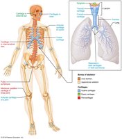
Types and Locations of Cartilage
The human skeleton initially consists of cartilage, which is gradually replaced by bone except in areas requiring flexibility. Cartilage is avascular and surrounded by the perichondrium, which supplies nutrients.
Hyaline Cartilage: Provides support, flexibility, and resilience; found in articular surfaces, costal cartilages, respiratory structures, and nasal cartilage.
Elastic Cartilage: Contains elastic fibers; found in the external ear and epiglottis.
Fibrocartilage: Contains thick collagen fibers; found in menisci of the knee and intervertebral discs.
Classification of Bones
Axial and Appendicular Skeleton
The 206 named bones of the human skeleton are divided into two groups:
Axial Skeleton: Skull, vertebral column, and rib cage (long axis of the body).
Appendicular Skeleton: Bones of the limbs and girdles attaching them to the axial skeleton.
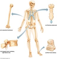
Bone Shapes
Long Bones: Longer than wide (e.g., humerus, femur).
Short Bones: Cube-shaped (e.g., wrist and ankle bones); sesamoid bones form within tendons (e.g., patella).
Flat Bones: Thin, flat, and slightly curved (e.g., sternum, scapulae, ribs, skull bones).
Irregular Bones: Complicated shapes (e.g., vertebrae, hip bones).
Bone Structure
Gross Anatomy of Bones
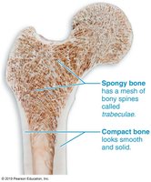
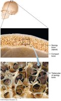
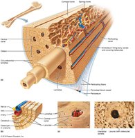
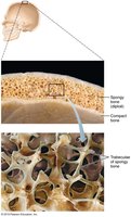
Compact Bone: Dense outer layer that appears smooth and solid.
Spongy Bone: Internal honeycomb of trabeculae filled with red or yellow marrow.
Structure of Short, Irregular, and Flat Bones
Thin plates of spongy bone (diploë) covered by compact bone.
Periosteum covers outside; endosteum covers inside.
No defined marrow cavity; marrow is scattered throughout spongy bone.
Hyaline cartilage covers areas involved in movable joints.
Structure of a Typical Long Bone
Diaphysis: Shaft; compact bone surrounding the medullary cavity (yellow marrow in adults).
Epiphyses: Bone ends; compact bone externally, spongy bone internally; articular cartilage covers joint surfaces.
Epiphyseal Line: Remnant of the growth plate between diaphysis and epiphysis.
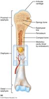
Bone Membranes
Periosteum: Double-layered membrane covering external surfaces except joints; outer fibrous layer (dense irregular connective tissue), inner osteogenic layer (bone-forming cells).
Endosteum: Delicate membrane covering internal bone surfaces, including trabeculae and canals.
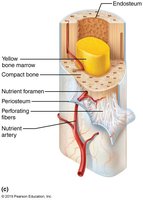
Bone Marrow
Red Marrow: Hematopoietic tissue found in trabecular cavities of spongy bone and diploë of flat bones; active in blood cell formation.
Yellow Marrow: Fat storage; can convert to red marrow if necessary (e.g., in anemia).
Bone Markings
Bone markings are anatomical features that serve as sites for muscle, ligament, and tendon attachment, joint formation, or passageways for blood vessels and nerves.
Name of Bone Marking | Description | Illustration |
|---|---|---|
Projection | Outward bulge of bone (e.g., tuberosity, crest, trochanter) | See image |
Depression | Bowl- or groove-like cut-out (e.g., fossa, groove) | See image |
Opening | Hole or canal (e.g., foramen, canal) | See image |
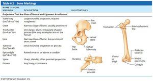
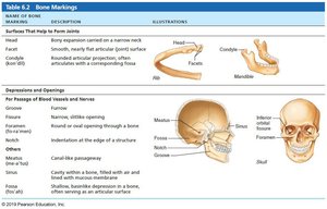
Microscopic Anatomy of Bone
Bone Cells
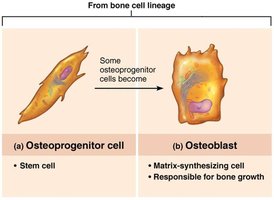
Osteogenic Cells: Stem cells in periosteum and endosteum; differentiate into osteoblasts or bone-lining cells.
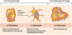
Osteoblasts: Bone-forming cells; secrete osteoid (unmineralized bone matrix).
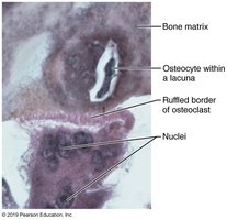
Osteocytes: Mature bone cells in lacunae; maintain bone matrix and act as stress sensors.
Bone-Lining Cells: Flat cells on bone surfaces; help maintain matrix.
Osteoclasts: Multinucleate cells derived from hematopoietic stem cells; responsible for bone resorption.
Compact Bone Structure
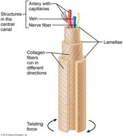
Osteon (Haversian System): Structural unit; elongated cylinder with concentric lamellae (rings of bone matrix).
Lamellae: Collagen fibers run in alternating directions in adjacent rings, providing strength and resistance to twisting.
Central (Haversian) Canal: Contains blood vessels and nerves.
Perforating (Volkmann's) Canals: Connect blood vessels and nerves of periosteum, medullary cavity, and central canal.
Lacunae: Small cavities containing osteocytes.
Canaliculi: Hairlike canals connecting lacunae, allowing communication and nutrient/waste exchange.
Interstitial and Circumferential Lamellae: Fill gaps between osteons and encircle the diaphysis, respectively.
Spongy Bone Structure
Spongy bone consists of trabeculae aligned along lines of stress, providing strength without excessive weight. Trabeculae contain irregularly arranged lamellae and osteocytes interconnected by canaliculi. Capillaries in the endosteum supply nutrients.
Chemical Composition of Bone
Organic and Inorganic Components
Organic: Includes bone cells and osteoid (ground substance and collagen fibers), providing tensile strength and flexibility.
Inorganic: Hydroxyapatites (mineral salts, mainly calcium phosphate) make up 65% of bone by mass, providing hardness and resistance to compression.
Bone is half as strong as steel in resisting compression and as strong as steel in resisting tension.
Bone Development (Ossification)
Types of Ossification
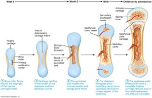
Endochondral Ossification: Bone forms by replacing hyaline cartilage; forms most of the skeleton below the skull (except clavicles).
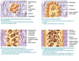
Intramembranous Ossification: Bone develops from fibrous membranes; forms flat bones of the skull and clavicles.
Steps of Intramembranous Ossification
Ossification centers form as mesenchymal cells become osteoblasts.
Osteoid is secreted and calcified.
Woven bone and periosteum form.
Lamellar bone replaces woven bone; red marrow appears.
Postnatal Bone Growth
Growth in Length (Interstitial Growth)
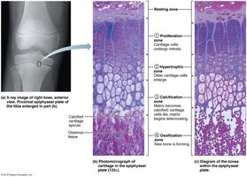
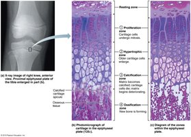
Long bones grow in length at the epiphyseal plate, which consists of five zones:
Resting (quiescent) zone
Proliferation (growth) zone
Hypertrophic zone
Calcification zone
Ossification (osteogenic) zone
Growth in Width (Appositional Growth)
Bones increase in thickness throughout life by appositional growth, where osteoblasts add bone matrix to the external surface and osteoclasts remove bone from the internal surface.
Bone Remodeling
Bone Deposit and Resorption
Bone Deposit: Osteoblasts secrete new bone matrix (osteoid seam), which is then mineralized.
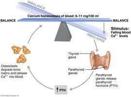
Bone Resorption: Osteoclasts break down bone matrix, releasing minerals into the blood.
Control of Bone Remodeling
Hormonal Regulation: Parathyroid hormone (PTH) stimulates osteoclasts to resorb bone when blood calcium is low; calcitonin (from the thyroid) can lower blood calcium at high doses.
Mechanical Stress: Bone remodeling is also regulated by mechanical forces and stress placed on bones.
Summary Table: Bone Cells and Functions
Cell Type | Origin | Function |
|---|---|---|
Osteogenic Cell | Mesenchymal stem cell | Bone stem cell; differentiates into osteoblasts |
Osteoblast | Osteogenic cell | Bone-forming cell; secretes osteoid |
Osteocyte | Osteoblast | Mature bone cell; maintains bone matrix |
Bone-lining Cell | Osteoblast | Maintains bone matrix on surfaces |
Osteoclast | Hematopoietic stem cell | Bone-resorbing cell |
Additional info: Understanding bone structure and function is essential for diagnosing and treating bone diseases such as osteoporosis, and for appreciating the dynamic nature of the skeletal system throughout life.
