 Back
BackBones and Skeletal Tissues: Structure, Function, and Classification
Study Guide - Smart Notes
 Tailored notes based on your materials, expanded with key definitions, examples, and context.
Bones and Skeletal Tissues
Introduction
The skeletal system forms the supporting framework of the human body, providing structure, protection, and facilitating movement. It is composed of bones, cartilages, ligaments, and connective tissues. Understanding the anatomy and physiology of bones and skeletal tissues is essential for comprehending how the body maintains its shape, protects vital organs, and enables locomotion.
The Skeletal System: Cartilages
Basic Structure, Types, and Locations of Cartilage
Skeletal cartilage is made of highly resilient, molded cartilage tissue that consists primarily of water, making it flexible and compressible.
Cartilage contains no blood vessels or nerves.
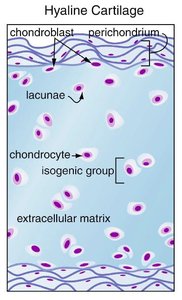
The perichondrium is a layer of dense connective tissue surrounding cartilage, providing nutrients and resisting outward expansion.
Chondrocytes are the cells found in small cavities (lacunae) within the extracellular matrix.

Types of Cartilage
Hyaline cartilage: Provides support, flexibility, and resilience; most abundant; found in articular (joints), costal (ribs), respiratory (larynx), and nasal cartilage.
Elastic cartilage: Similar to hyaline but contains elastic fibers; found in the external ear and epiglottis.
Fibrocartilage: Contains thick collagen fibers for tensile strength; found in menisci of the knee and intervertebral discs.
Growth of Cartilage
Appositional growth: Cartilage-forming cells in the perichondrium secrete new matrix on the surface of existing cartilage.
Interstitial growth: Chondrocytes within lacunae divide and secrete new matrix, expanding cartilage from within.
Calcification: Occurs during normal bone growth in youth and sometimes in old age; calcified cartilage is not bone.

Functions of the Skeletal System
Major Functions of Bones
Support: Provides a framework for the body and supports soft organs.
Protection: Protects vital organs such as the brain, spinal cord, and thoracic organs.
Movement: Acts as levers for muscles to produce movement.
Mineral and growth factor storage: Stores calcium, phosphorus, and growth factors.
Blood cell formation (hematopoiesis): Occurs in red marrow cavities of certain bones.
Triglyceride (fat) storage: Fat is stored in yellow marrow cavities for energy.
Hormone production: Osteocalcin is secreted by bones to regulate insulin secretion, glucose levels, and metabolism.
Classification of Bones
Axial vs. Appendicular Skeleton
Axial skeleton: Forms the long axis of the body (skull, vertebral column, rib cage).
Appendicular skeleton: Includes bones of the upper and lower limbs and girdles attaching limbs to the axial skeleton.

Classification by Shape
Long bones: Longer than they are wide (e.g., humerus, femur).
Short bones: Cube-shaped (e.g., carpal bones of the wrist).
Flat bones: Thin, flat, and often curved (e.g., sternum, skull, ribs, scapula).
Irregular bones: Complicated shapes (e.g., vertebrae, pelvic bones).
Sutural bones: Small, irregular bones found between the flat bones of the skull.
Sesamoid bones: Small and flat, develop inside tendons (e.g., patella).


Bone Markings
Types and Functions of Bone Markings
Bone markings are surface features that serve as sites of muscle, ligament, and tendon attachment, as well as passages for nerves and blood vessels.
General Description | Anatomical Term | Definition |
|---|---|---|
Projection | Process, Ramus, Trochanter, Tuberosity, Crest, Line, Spine | Outward bulges, often for muscle or ligament attachment |
Depression | Fossa, Sulcus | Bowl- or groove-like cut-outs for passage of vessels/nerves or joint formation |
Opening | Foramen, Canal, Meatus, Sinus | Holes or canals for passage of blood vessels and nerves |


Bone Structure
Gross Anatomy of Bones
Long bones have a shaft (diaphysis), bone ends (epiphyses), and membranes.
Diaphysis: Tubular shaft with compact bone surrounding a medullary cavity (yellow marrow in adults).
Epiphyses: Ends of long bones with compact bone externally and spongy bone internally; articular cartilage covers joint surfaces.
Metaphysis: Region where diaphysis and epiphysis meet; contains the epiphyseal line (remnant of growth plate).

Compact vs. Spongy Bone
Compact bone: Dense outer layer that appears smooth and solid.
Spongy bone: Honeycomb structure of trabeculae with open spaces filled with red or yellow marrow.

Microscopic Anatomy of Bone
Osteon (Haversian system): Structural unit of compact bone; consists of concentric lamellae around a central canal.
Lamellae: Rings of bone matrix with alternating collagen fiber orientation for strength.
Canaliculi: Small canals connecting lacunae, allowing communication and nutrient/waste exchange between osteocytes.
Interstitial and circumferential lamellae: Fill gaps between osteons and encircle the diaphysis, respectively.
Bone Cells
Osteogenic (osteoprogenitor) cells: Stem cells in periosteum and endosteum; differentiate into osteoblasts.
Osteoblasts: Bone-forming cells that secrete osteoid (unmineralized bone matrix).
Osteocytes: Mature bone cells in lacunae; maintain bone matrix and act as mechanosensors.
Bone-lining cells: Flat cells on bone surfaces; help maintain matrix.
Osteoclasts: Multinucleate cells responsible for bone resorption; derived from hematopoietic stem cells.
Chemical Composition of Bone
Organic and Inorganic Components
Organic: Osteogenic cells, osteoblasts, osteocytes, bone-lining cells, osteoclasts, and osteoid (collagen and ground substance).
Inorganic: Hydroxyapatite (calcium phosphate crystals) makes up 65% of bone by mass, providing hardness and resistance to compression.
Key Equation:
This reaction forms hydroxyapatite, the main mineral component of bone.
Summary Table: Types of Bone Cells
Cell Type | Function |
|---|---|
Osteogenic cells | Stem cells; differentiate into osteoblasts |
Osteoblasts | Bone formation; secrete osteoid |
Osteocytes | Maintain bone matrix; mechanosensors |
Bone-lining cells | Maintain bone matrix on surfaces |
Osteoclasts | Bone resorption (breakdown) |
Conclusion
The skeletal system is a dynamic organ system essential for support, movement, protection, and metabolic functions. Bones are classified by shape and structure, and their composition allows them to be both strong and flexible. Understanding the anatomy and physiology of bones and skeletal tissues is foundational for further study in human anatomy and physiology.
