 Back
BackBones and Skeletal Tissues: Structure, Function, and Clinical Aspects
Study Guide - Smart Notes
 Tailored notes based on your materials, expanded with key definitions, examples, and context.
Bones and Skeletal Tissues
Cartilage: Basic Structure, Types, and Locations
Cartilage is a resilient, avascular connective tissue found throughout the body, especially in joints, the respiratory tract, and certain skeletal structures. It is surrounded by a dense connective tissue layer called the perichondrium, which provides nutrients and resists outward expansion.
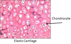
Chondrocytes are the main cell type, residing in lacunae within the extracellular matrix.
Cartilage contains no blood vessels or nerves.
Types of cartilage:
Hyaline cartilage: Most abundant; provides support, flexibility, and resilience. Found in articular surfaces, costal cartilage, respiratory tract, and nose.
Elastic cartilage: Similar to hyaline but contains elastic fibers, allowing flexibility. Found in the external ear and epiglottis.
Fibrocartilage: Contains thick collagen fibers, providing great tensile strength. Found in intervertebral discs, menisci of the knee, and pubic symphysis.
Growth of Cartilage
Cartilage grows by two mechanisms:
Appositional growth: Cells in the perichondrium secrete new matrix on the external surface.
Interstitial growth: Chondrocytes divide and secrete new matrix from within, expanding the cartilage.
Calcification of cartilage occurs during normal bone growth (youth and old age), but calcified cartilage is not bone.
Classification of Bones
Axial and Appendicular Skeleton
The human skeleton consists of 206 named bones, divided into two main groups:
Axial skeleton: Skull, vertebral column, and rib cage (long axis of the body).
Appendicular skeleton: Bones of the limbs and girdles attaching them to the axial skeleton.
Classification of Bones by Shape
Long bones: Longer than wide (e.g., humerus, femur).
Short bones: Cube-shaped (e.g., wrist and ankle bones); includes sesamoid bones (e.g., patella).
Flat bones: Thin, flat, and slightly curved (e.g., sternum, scapulae, ribs, skull bones).
Irregular bones: Complicated shapes (e.g., vertebrae, coxal bones).
Functions of Bones
Bones serve several critical functions in the body:
Support: Framework for the body and soft organs.
Protection: Shields the brain, spinal cord, and vital organs.
Movement: Act as levers for muscle action.
Mineral and growth factor storage: Reservoir for calcium, phosphorus, and growth factors.
Blood cell formation (hematopoiesis): Occurs in red marrow cavities.
Triglyceride (fat) storage: Stored in bone cavities as an energy source.
Hormone production: Osteocalcin regulates bone formation and metabolism.
Bone Structure
Gross Anatomy: Compact and Spongy Bone
Bones have two main textures:
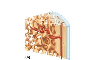
Compact bone: Dense outer layer; smooth and solid.
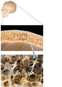
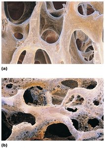
Spongy bone (cancellous or trabecular): Honeycomb of trabeculae deep to compact bone.
Structure of Short, Irregular, and Flat Bones
Thin plates of spongy bone covered by compact bone.
Sandwiched between periosteum (outer) and endosteum (inner).
No shaft or epiphyses; bone marrow throughout spongy bone, but no marrow cavity.
Hyaline cartilage covers articular surfaces at joints.
Structure of a Typical Long Bone
Diaphysis: Tubular shaft; compact bone surrounds the medullary cavity (contains yellow marrow).
Epiphyses: Bone ends; external compact bone, internal spongy bone, articular cartilage covers joint surfaces.
Epiphyseal line: Remnant of the growth plate between diaphysis and epiphysis.

Membranes: Periosteum and Endosteum
Periosteum: Double-layered membrane covering external bone surfaces (except joints); outer fibrous layer (dense irregular connective tissue) and inner osteogenic layer (contains osteogenic cells).
Endosteum: Delicate membrane covering internal bone surfaces, including trabeculae and canals; contains osteogenic cells.
Hematopoietic Tissue in Bones
Red marrow: Found in trabecular cavities of spongy bone and diploë of flat bones; active in hematopoiesis.
In adults, red marrow is limited to heads of femur and humerus, diploë, and some irregular bones.
Yellow marrow can convert to red marrow if necessary.
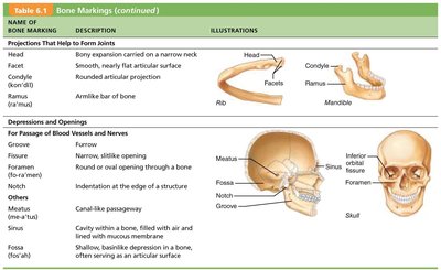
Bone Markings
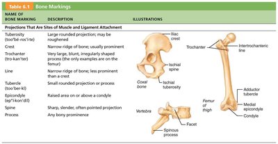
Bone markings are anatomical features that serve as sites for muscle, ligament, and tendon attachment, joint surfaces, or conduits for blood vessels and nerves. They are classified as projections, depressions, or openings.
Name of Bone Marking | Description | Illustration |
|---|---|---|
Process | Any bony prominence | Various bones |
Trochanter | Very large, blunt, irregularly shaped process | Femur |
Crest | Narrow ridge of bone; usually prominent | Iliac crest |
Condyle | Rounded articular projection | Mandible |
Foramen | Round or oval opening through a bone | Skull |
Fossa | Shallow basin-like depression | Skull |
Meatus | Canal-like passageway | Skull |
Sinus | Cavity within a bone, filled with air and lined with mucous membrane | Skull |
Microscopic Anatomy of Bone
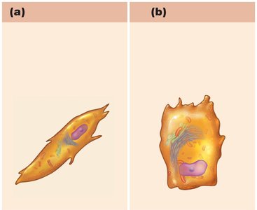
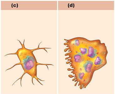
Cells of Bone Tissue
Bone tissue contains five major cell types, all derived from the same basic cell lineage:
Osteogenic cells: Stem cells in periosteum and endosteum; differentiate into osteoblasts or bone lining cells.
Osteoblasts: Bone-forming cells; secrete unmineralized bone matrix (osteoid).
Osteocytes: Mature bone cells in lacunae; maintain bone matrix and act as stress sensors.
Bone lining cells: Flat cells on bone surfaces; help maintain matrix.
Osteoclasts: Multinucleate cells derived from hematopoietic stem cells; responsible for bone resorption.
Compact Bone (Lamellar Bone)
The structural unit of compact bone is the osteon (Haversian system), an elongated cylinder parallel to the long axis of the bone. Each osteon consists of concentric lamellae of bone matrix, with collagen fibers running in alternating directions to resist twisting forces.
Spongy Bone
Spongy bone appears poorly organized but is highly adaptive. Its trabeculae align along lines of stress, providing structural support without the weight of compact bone. Trabeculae contain irregularly arranged lamellae and osteocytes interconnected by canaliculi.
Bone Development and Growth
Ossification (Osteogenesis)
Ossification is the process of bone tissue formation, beginning in the second month of development and continuing through early adulthood. It includes:
Formation of the bony skeleton
Postnatal bone growth (length and width)
Bone remodeling and repair (lifelong)
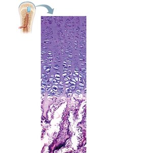
Interstitial Growth: Growth in Length of Long Bones
Long bones grow in length at the epiphyseal plate through the following zones:
Resting zone: Inactive cartilage cells
Proliferation zone: Chondrocytes undergo mitosis
Hypertrophic zone: Older cartilage cells enlarge
Calcification zone: Matrix calcifies, cartilage cells die, blood vessels invade
Ossification zone: New bone forms
Appositional Growth: Growth in Width
Bones increase in diameter through appositional growth, where osteoblasts beneath the periosteum secrete bone matrix on the external surface, while osteoclasts remove bone from the endosteal surface. This process results in thicker, stronger bones.
Hormonal Regulation of Bone Growth
Growth hormone: Stimulates epiphyseal plate activity in children.
Thyroid hormone: Modulates growth hormone activity for proper proportions.
Sex hormones (testosterone, estrogens): Promote growth spurts and induce epiphyseal plate closure at puberty.
Imbalances can cause abnormal skeletal growth.
Bone Homeostasis and Remodeling
Bone Remodeling
Bone remodeling is a continuous process involving bone deposit by osteoblasts and bone resorption by osteoclasts. Remodeling occurs at the surfaces of both periosteum and endosteum, maintaining bone strength and mineral homeostasis.
Importance of Calcium
Calcium is essential for nerve impulse transmission, muscle contraction, blood coagulation, gland and nerve cell secretion, and cell division. The body contains 1200–1400 grams of calcium, with 99% stored in bones. Blood calcium levels are tightly regulated (9–11 mg/dl).
Hormonal Control of Blood Calcium
Parathyroid hormone (PTH): Increases blood calcium by stimulating bone resorption.
Calcitonin: Lowers blood calcium levels temporarily in high doses.
Other hormones: Leptin (inhibits osteoblasts), serotonin (interferes with osteoblast activity).
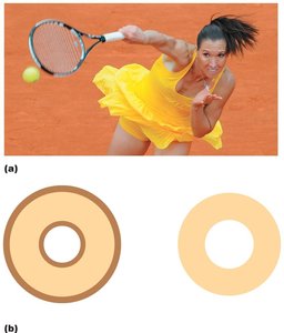
Response to Mechanical Stress
Bones adapt to the stresses placed upon them. Mechanical stress stimulates bone remodeling, making bones thicker and stronger where needed. For example, the serving arm of a tennis player has a thicker humerus due to repeated stress.
Bone Repair and Fractures
Fracture Classification
Fractures are classified by:
Position of bone ends (nondisplaced/displaced)
Completeness of break (complete/incomplete)
Whether skin is penetrated (open/closed)
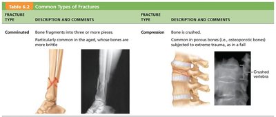
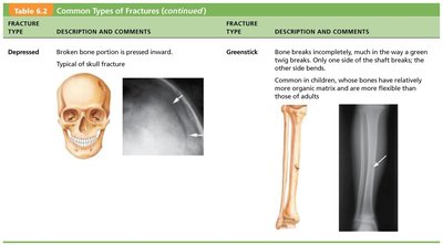
Common Types of Fractures
Fracture Type | Description and Comments |
|---|---|
Comminuted | Bone fragments into three or more pieces; common in aged, brittle bones. |
Compression | Bone is crushed; common in porous bones subjected to trauma. |
Spiral | Ragged break from excessive twisting forces; common sports fracture. |
Epiphyseal | Epiphysis separates from diaphysis along epiphyseal plate; occurs when cartilage cells are dying. |
Depressed | Broken bone portion pressed inward; typical of skull fracture. |
Greenstick | Bone breaks incompletely; common in children. |

Fracture Treatment and Repair
Reduction: Realignment of broken bone ends (closed or open reduction).
Immobilization: By cast or traction for healing; depends on severity, bone, and patient age.
Homeostatic Imbalances of Bone
Osteomalacia and Rickets
Osteomalacia: Poorly mineralized, soft, weak bones; pain upon bearing weight; caused by inadequate calcium or vitamin D.
Rickets: Osteomalacia in children; causes bowed legs and bone deformities.
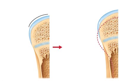
Osteoporosis
Bone resorption outpaces deposit; spongy bone of spine and femur most susceptible.
Common in aged, postmenopausal women; risk factors include petite body form, poor diet, smoking, immobility, and hormone-related conditions.
Treatments: Calcium, vitamin D, exercise, hormone replacement therapy (controversial).
Prevention: Adequate calcium, reduced carbonated beverages/alcohol, weight-bearing exercise.
Age-Related Changes in Bone
Bone formation exceeds resorption in children/adolescents; balanced in young adults; resorption exceeds formation with age.
Bone mass, mineralization, and healing ability decrease with age, except in skull bones.
Genetics play a major role in bone density and osteoporosis risk.
