 Back
BackBones and Skeletal Tissues: Structure, Function, and Clinical Aspects
Study Guide - Smart Notes
 Tailored notes based on your materials, expanded with key definitions, examples, and context.
Bones and Skeletal Tissues
Cartilage: Basic Structure, Types, and Locations
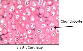
Cartilage is a resilient, avascular connective tissue found throughout the body, especially in joints, the respiratory tract, and certain skeletal structures. It is surrounded by a dense connective tissue layer called the perichondrium, which provides nutrients and resists outward expansion.
Chondrocytes: The primary cells of cartilage, located in lacunae within the extracellular matrix.
Types of Cartilage:
Hyaline cartilage: Most abundant; provides support and flexibility. Found in articular surfaces, costal cartilages, respiratory tract, and nose.
Elastic cartilage: Contains elastic fibers for flexibility; found in the external ear and epiglottis.
Fibrocartilage: Contains thick collagen fibers for tensile strength; found in intervertebral discs, menisci, and pubic symphysis.
Growth of Cartilage:
Appositional growth: New matrix is secreted on the external surface.
Interstitial growth: Chondrocytes divide and secrete new matrix from within.
Calcification: Occurs during bone growth or aging, but calcified cartilage is not bone.
Classification of Bones
Axial and Appendicular Skeleton
The human skeleton consists of 206 named bones, divided into two main groups:
Axial skeleton: Skull, vertebral column, and rib cage (long axis of the body).
Appendicular skeleton: Bones of the limbs and girdles attaching them to the axial skeleton.
Classification by Shape
Long bones: Longer than wide (e.g., humerus, femur).
Short bones: Cube-shaped (e.g., wrist and ankle bones); includes sesamoid bones (e.g., patella).
Flat bones: Thin, flat, and slightly curved (e.g., sternum, scapulae, ribs, skull bones).
Irregular bones: Complicated shapes (e.g., vertebrae, coxal bones).
Functions of Bones
Bones serve several critical functions in the body:
Support: Framework for the body and soft organs.
Protection: Shields the brain, spinal cord, and vital organs.
Movement: Act as levers for muscle action.
Mineral and growth factor storage: Reservoir for calcium, phosphorus, and growth factors.
Blood cell formation (hematopoiesis): Occurs in red marrow cavities.
Triglyceride storage: Fat stored in bone cavities as an energy source.
Hormone production: Osteocalcin regulates bone formation and metabolism.
Bone Structure
Gross Anatomy
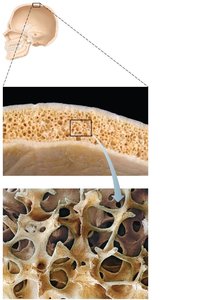
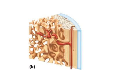
Compact bone: Dense outer layer; smooth and solid.
Spongy bone (cancellous or trabecular): Honeycomb of trabeculae deep to compact bone.
Structure of Long Bones
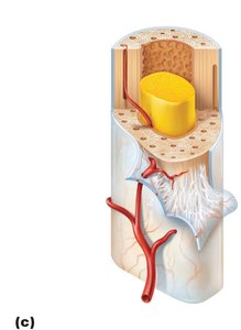
Diaphysis: Tubular shaft; compact bone surrounds the medullary cavity (contains yellow marrow).
Epiphyses: Bone ends; external compact bone, internal spongy bone, articular cartilage covers joint surfaces.
Epiphyseal line: Remnant of the growth plate between diaphysis and epiphysis.
Membranes
Periosteum: Double-layered membrane covering external bone (except joints); outer fibrous layer and inner osteogenic layer.
Endosteum: Delicate membrane covering internal bone surfaces, including trabeculae and canals.
Hematopoietic Tissue in Bones
Red marrow: Found in trabecular cavities of spongy bone and diploë of flat bones; active in hematopoiesis.
Yellow marrow: Fat storage; can convert to red marrow if necessary.
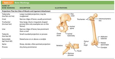
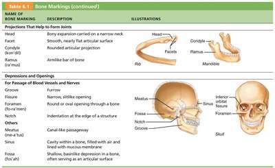
Bone Markings
Bone markings are structural features adapted for muscle, ligament, and tendon attachment, joint formation, and passage of blood vessels and nerves.
Name | Description | Illustration |
|---|---|---|
Process | Any bony prominence | Vertebra |
Trochanter | Large, blunt, irregular surface | Femur |
Crest | Narrow ridge of bone | Iliac crest |
Condyle | Rounded articular projection | Mandible |
Foramen | Round or oval opening | Skull |
Fossa | Shallow depression | Skull |
Meatus | Canal-like passageway | Skull |
Sinus | Cavity within a bone | Skull |
Microscopic Anatomy of Bone
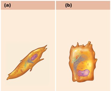
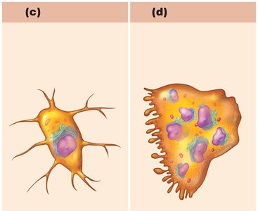
Bone Cells
Osteogenic cells: Stem cells in periosteum and endosteum; differentiate into osteoblasts or bone lining cells.
Osteoblasts: Bone-forming cells; secrete osteoid (unmineralized bone matrix).
Osteocytes: Mature bone cells in lacunae; maintain bone matrix and act as stress sensors.
Bone lining cells: Flat cells on bone surfaces; help maintain matrix.
Osteoclasts: Multinucleate cells for bone resorption; derived from hematopoietic stem cells.
Compact Bone (Lamellar Bone)
Osteon (Haversian system): Structural unit; elongated cylinders parallel to bone's long axis.
Lamellae: Concentric rings of bone matrix; collagen fibers run in alternating directions to resist twisting.
Central canal: Contains blood vessels and nerves.
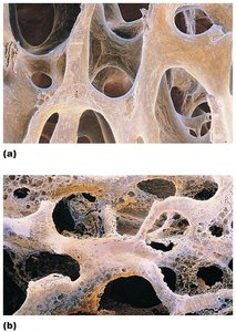
Spongy Bone
Trabeculae: Irregularly arranged lamellae; align along lines of stress for strength.
No osteons; nutrients supplied by capillaries in endosteum.
Bone Development and Growth
Ossification (Osteogenesis)
The process of bone tissue formation, including the formation of the bony skeleton, postnatal bone growth, and bone remodeling and repair.
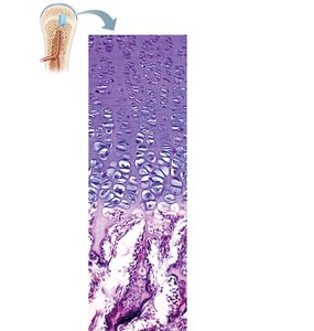
Growth in Length of Long Bones
Occurs at the epiphyseal plate through proliferation, hypertrophy, calcification, and ossification zones.
Epiphyseal plate closure marks the end of longitudinal growth (females ~18 years, males ~21 years).
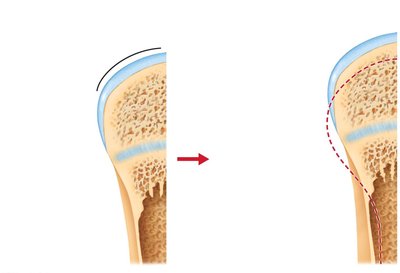
Appositional Growth (Width)
Osteoblasts add bone matrix to the external surface; osteoclasts remove bone from the internal surface.
Results in thicker, stronger bones.
Hormonal Regulation of Bone Growth
Growth hormone: Stimulates epiphyseal plate activity.
Thyroid hormone: Modulates growth hormone effects.
Sex hormones: Promote growth spurts and induce epiphyseal plate closure at puberty.
Bone Homeostasis
Bone Remodeling
Continuous process of bone deposit and resorption at periosteum and endosteum surfaces.
Remodeling units consist of osteoblasts and osteoclasts.
Calcium Homeostasis
Calcium is essential for nerve transmission, muscle contraction, blood coagulation, and more.
Blood calcium is regulated by parathyroid hormone (PTH) (increases blood Ca2+ by stimulating bone resorption) and calcitonin (lowers blood Ca2+ in high doses).
Other hormones: Leptin (inhibits osteoblasts), serotonin (interferes with osteoblast activity).
Response to Mechanical Stress
Bones adapt to the stresses placed upon them; thickest where stress is greatest.
Mechanical stress determines where remodeling occurs.
Bone Repair
Fracture Classification
Position: Nondisplaced (normal alignment) vs. displaced (misaligned).
Completeness: Complete (all the way through) vs. incomplete.
Skin penetration: Open (compound) vs. closed (simple).
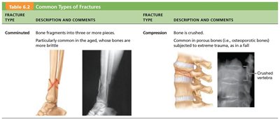
Fracture Type | Description |
|---|---|
Comminuted | Bone fragments into three or more pieces; common in aged, brittle bones. |
Compression | Bone is crushed; common in porous bones (e.g., vertebrae). |
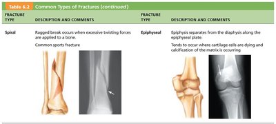
Spiral | Ragged break from excessive twisting; common sports fracture. |
Epiphyseal | Epiphysis separates from diaphysis along epiphyseal plate. |
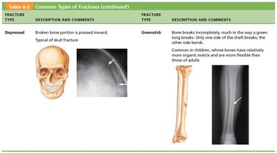
Depressed | Broken bone portion pressed inward; typical of skull fracture. |
Greenstick | Bone breaks incompletely; common in children. |
Fracture Treatment and Repair
Reduction: Realignment of bone ends (closed or open).
Immobilization: Cast or traction for healing; depends on severity, bone, and patient age.
Homeostatic Imbalances of Bone
Osteomalacia and Rickets
Osteomalacia: Poorly mineralized, soft, weak bones; pain on weight-bearing.
Rickets: Osteomalacia in children; bowed legs, bone deformities, enlarged bone ends. Caused by vitamin D deficiency or insufficient calcium.
Osteoporosis
Bone resorption outpaces deposit; spongy bone of spine and femur most affected.
Common in postmenopausal women; risk factors include petite body, poor diet, smoking, immobility, hormone imbalances.
Treatment: Calcium, vitamin D, exercise, hormone therapy (controversial).
Prevention: Adequate calcium, reduced alcohol/carbonated drinks, weight-bearing exercise.
Age-Related Changes in Bone
Bone formation exceeds resorption in children/adolescents; balanced in young adults; resorption exceeds formation with age.
Bone mass and healing ability decrease with age, except in the skull.
