 Back
BackBones and Skeletal Tissues: Structure, Function, and Growth
Study Guide - Smart Notes
 Tailored notes based on your materials, expanded with key definitions, examples, and context.
Bones and Skeletal Tissues
Overview of Cartilages in the Body
Cartilage is a resilient and smooth elastic tissue, a major component of the skeletal system. It is found in various locations throughout the body, providing support and flexibility where needed.
Locations: Cartilage is present in the external ear, nose, joints, rib cage, intervertebral discs, and respiratory structures.
Types: The three main types of cartilage are hyaline, elastic, and fibrocartilage.
Function: Cartilage cushions joints, supports soft tissues, and forms the embryonic skeleton.
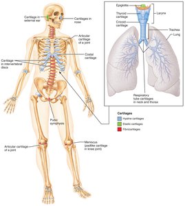
Types of Cartilage Tissue
Cartilage tissue is classified based on its composition and function. Each type has unique structural features and locations in the body.
Hyaline Cartilage: Most abundant; provides support with flexibility. Found in articular surfaces, costal cartilages, nose, and respiratory passages.
Elastic Cartilage: Contains elastic fibers; maintains shape and flexibility. Found in the external ear and epiglottis.
Fibrocartilage: Contains thick collagen fibers; resists compression and tension. Found in intervertebral discs and pubic symphysis.
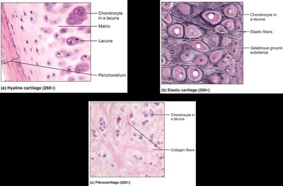
Growth of Cartilage
Cartilage grows by two mechanisms, both essential during development and repair.
Appositional Growth: Chondroblasts in the perichondrium produce new cartilage on the surface.
Interstitial Growth: Chondrocytes within the cartilage divide and secrete new matrix internally.
Growth Limitation: Cartilage growth ceases when the skeleton stops growing.
Tissues in Bone
Bones are complex organs composed of several tissue types, each contributing to their function and structure.
Bone Connective Tissue: Dominates bone structure.
Nervous Tissue: Present in bone for sensory and regulatory functions.
Blood Connective Tissue: Supplies nutrients and removes waste.
Cartilage: Found in articular surfaces.
Epithelial Tissue: Lines blood vessels within bone.
Functions of Bones
Bones serve multiple essential functions in the human body.
Support: Provide a rigid framework for the body.
Movement: Act as levers for muscles.
Protection: Shield vital organs (e.g., brain, heart).
Mineral Storage: Store calcium and phosphate.
Blood Cell Formation: Red marrow produces blood cells.
Energy Metabolism: Osteoblasts secrete osteocalcin, influencing metabolism.
Classification of Bones
Bones are classified by their shape and structure, which relates to their function.
Long Bones: Longer than wide; have a shaft and two ends (e.g., femur).
Short Bones: Cube-shaped (e.g., carpals).
Flat Bones: Thin, flattened, often curved (e.g., sternum).
Irregular Bones: Complex shapes (e.g., vertebrae).
Gross Anatomy of Bones
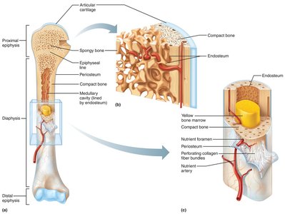
Bones have two main structural types: compact and spongy bone.
Compact Bone: Dense outer layer providing strength.
Spongy (Cancellous) Bone: Internal network of trabeculae, filled with marrow.
Structure of a Typical Long Bone
Long bones have distinct anatomical regions and features.
Diaphysis: Shaft of the bone.
Epiphyses: Ends of the bone.
Medullary Cavity: Central cavity containing yellow marrow.
Membranes: Periosteum (outer), endosteum (inner), and Sharpey's fibers (collagen bundles).
Blood Vessels: Bones are highly vascularized.
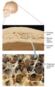
Structure of a Flat Bone
Flat bones consist of two layers of compact bone enclosing a layer of spongy bone (diploë).
Bone Markings
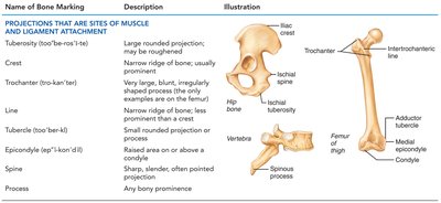
Bone markings are features on the surface of bones that reflect stresses and serve as sites for muscle attachment, joint formation, or passage of vessels and nerves.
Projections: For muscle and ligament attachment (e.g., tuberosity, crest, trochanter).
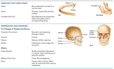
Surfaces: Form joints (e.g., head, facet, condyle).
Depressions and Openings: Allow passage of vessels and nerves (e.g., foramen, groove, fissure).
Name of Bone Marking | Description | Illustration |
|---|---|---|
Tuberosity | Large rounded projection; may be roughened | Hip bone |
Crest | Narrow ridge of bone; usually prominent | Hip bone |
Trochanter | Very large, blunt, irregularly shaped process | Femur |
Line | Narrow ridge of bone; less prominent than crest | Femur |
Tubercle | Small rounded projection or process | Femur |
Epicondyle | Raised area on or above a condyle | Femur |
Spine | Sharp, slender, often pointed projection | Spinous process |
Process | Any bony prominence | Any bone |
Surfaces That Form Joints | Description | Illustration |
|---|---|---|
Head | Bony expansion carried on a narrow neck | Head of femur |
Facet | Smooth, nearly flat articular surface | Facet of rib |
Condyle | Rounded articular projection | Condyle of mandible |
Depressions and Openings | Description | Illustration |
|---|---|---|
Foramen | Round or oval opening through a bone | Skull |
Groove | Furrow | Skull |
Fissure | Narrow, slitlike opening | Skull |
Notch | Indentation at the edge of a structure | Skull |
Fossa | Shallow basinlike depression | Skull |
Meatus | Canal-like passageway | Skull |
Sinus | Cavity within a bone, filled with air and lined with mucosa | Skull |
Microscopic Structure of Compact Bone
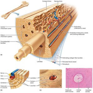
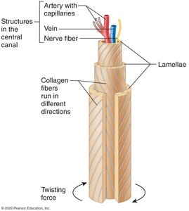
Compact bone is organized into osteons, which are cylindrical structures that provide support and house blood vessels and nerves.
Osteons (Haversian Systems): Resemble tree rings in cross-section.
Lamellae: Concentric layers of bone matrix.
Central Canal: Contains blood vessels and nerves.
Perforating Canals: Connect central canals.
Canaliculi: Small channels connecting osteocytes.
Microscopic Structure of Spongy Bone
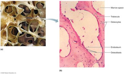
Spongy bone is less dense and consists of trabeculae, which are small, beam-like structures containing lamellae and osteocytes.
Trabeculae: Network of bone beams.
Marrow Spaces: Filled with red or yellow marrow.
Osteocytes: Bone cells within trabeculae.
Endosteum: Membrane lining internal bone surfaces.
Organization of Cartilage Within the Epiphyseal Plate
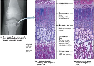
The epiphyseal plate is a region of growing long bones, organized into distinct zones that facilitate bone elongation.
Resting Zone: Quiescent cartilage cells.
Proliferation Zone: Rapidly dividing cartilage cells.
Hypertrophic Zone: Enlarged cartilage cells.
Calcification Zone: Matrix becomes calcified; cells die.
Ossification Zone: New bone forms.
Remodeling of Spongy Bone
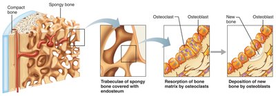
Bone remodeling is a continuous process involving the resorption of old bone and deposition of new bone by osteoclasts and osteoblasts.
Osteoclasts: Cells that break down bone matrix.
Osteoblasts: Cells that build new bone matrix.
Remodeling: Maintains bone strength and mineral homeostasis.
Key Equations and Concepts
Bone Matrix Composition:
Osteon Structure:
Summary Table: Bone Types and Features
Bone Type | Shape | Example |
|---|---|---|
Long Bone | Longer than wide | Femur |
Short Bone | Cube-shaped | Carpals |
Flat Bone | Thin, flattened, curved | Sternum |
Irregular Bone | Complex shape | Vertebrae |
Additional info:
Bone remodeling is influenced by mechanical stress and hormonal regulation.
Osteocalcin, secreted by osteoblasts, plays a role in energy metabolism and regulation of blood glucose.
