 Back
Bones and Skeletal Tissues: Structure, Function, and Physiology
Study Guide - Smart Notes
 Tailored notes based on your materials, expanded with key definitions, examples, and context.
Chapter 6: Bones & Skeletal Tissues
Overview of the Skeletal System
The skeletal system is a complex framework that provides structural support, protection, and facilitates movement. It consists of bones, bone marrow, cartilages, ligaments, and joints, with 206 bones in the adult human body.
Support: Provides a framework for organs and soft tissues.
Protection: Encases vital organs such as the brain, spinal cord, heart, and lungs.
Anchorage: Bones act as levers for muscles, enabling movement at joints.
Mineral Storage: Stores calcium (Ca2+) and phosphate (PO43-).
Blood Cell Formation: Bone marrow produces red blood cells, white blood cells, and platelets.
Fat Storage: Yellow bone marrow stores triglycerides for energy.
Hormone Production: Osteocalcin regulates bone formation and metabolism.
Cartilage Tissue: Structure and Types
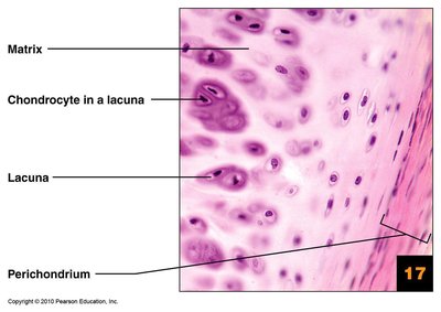
Cartilage is a specialized connective tissue that provides support and cushioning. It is flexible, consists mostly of water, and resists compression. Chondrocytes are the main cells, located in lacunae within a gel-like matrix rich in collagen fibers. Cartilage is avascular and surrounded by the perichondrium, a dense irregular connective tissue layer.
Hyaline Cartilage: Most common; contains only collagen fibers. Provides support with flexibility and resilience. Found in articular, costal, respiratory, and nasal regions.
Elastic Cartilage: Contains elastic fibers in addition to collagen, making it highly flexible. Found in the external ear and epiglottis.
Fibrocartilage: Contains thick collagen fibers, making it the strongest type. Resists compression; found in intervertebral discs and menisci.
Classification of Bones
Bone Shape Classes and Skeletal Divisions
Bones are classified by their shape and location within the skeleton. The axial skeleton provides support and protection, while the appendicular skeleton facilitates movement.
Long Bones: Longer than wide, with a shaft and two ends (e.g., femur, humerus).
Short Bones: Cube-shaped (e.g., wrist and ankle bones).
Sesamoid Bones: Small, round bones embedded within tendons (e.g., patella).
Flat Bones: Thin, flattened, and slightly curved (e.g., sternum, ribs, cranial bones).
Irregular Bones: Complex shapes (e.g., vertebrae, hip bones).
Gross Anatomy of Bones
Structure of Long Bones
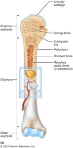
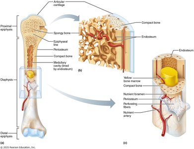
Long bones have a distinct anatomy, including a diaphysis (shaft), epiphyses (ends), and metaphysis (region between shaft and ends). The diaphysis contains compact bone and a medullary cavity filled with yellow marrow in adults. Epiphyses contain spongy bone and are covered by articular cartilage.
Diaphysis: Thick compact bone surrounds the medullary cavity.
Epiphyses: Compact bone outside, spongy bone inside, covered by articular cartilage.
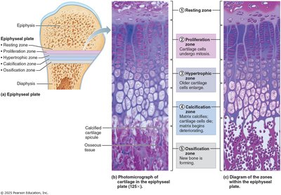
Metaphysis: Contains the epiphyseal plate (growth plate) in growing bones, replaced by the epiphyseal line in adults.
Membranes of Bone
Bones are covered externally by the periosteum and internally lined by the endosteum. The periosteum has a fibrous layer for protection and an osteogenic layer for bone growth. Perforating fibers anchor tendons and ligaments.
Structure of Short, Flat, and Irregular Bones
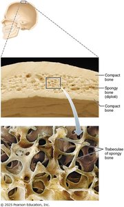
These bones lack a diaphysis and medullary cavity. They consist of thin plates of spongy bone (diploë) sandwiched between compact bone, with bone marrow present. Periosteum covers the outside, and endosteum lines the inside.
Bone Marrow: Red vs. Yellow
Bone marrow is found within the cavities of bones and is essential for hematopoiesis and fat storage.
Red Marrow: Hematopoietic tissue; produces RBCs, WBCs, and platelets. Located in spongy bone of flat bones and epiphyses of long bones in adults; nearly all marrow is red in infants.
Yellow Marrow: Primarily adipose tissue; found in the medullary cavity of adult long bones. Can convert to red marrow if needed.
Bone Markings
Bones display various markings, including projections, surfaces, depressions, and openings, which serve as sites for muscle attachment, joint formation, and passage of blood vessels and nerves.
Microscopic Anatomy of Bone
Bone Cells
Bony tissue contains several cell types, each with distinct functions:
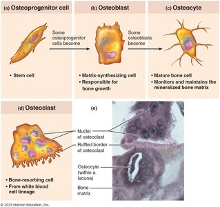
Osteoprogenitor (osteogenic) cells: Stem cells that differentiate into osteoblasts.
Osteoblasts: Matrix-synthesizing cells responsible for bone growth; secrete osteoid.
Osteocytes: Mature bone cells that monitor and maintain the mineralized bone matrix.
Osteoclasts: Bone-resorbing cells derived from white blood cell lineage; break down bone tissue.
Compact Bone: The Osteon
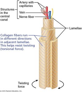
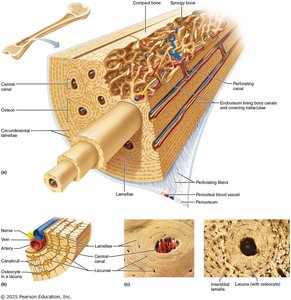
Compact bone is organized into osteons (Haversian systems), which are cylindrical structures containing concentric lamellae, a central canal, lacunae, and canaliculi. Collagen fibers in adjacent lamellae run in different directions, providing resistance to torsional forces.
Spongy Bone
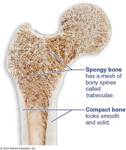
Spongy bone consists of a network of trabeculae, which provide structural support and house bone marrow. It is lighter than compact bone and found primarily in the epiphyses of long bones and within flat bones.
Chemical Composition of Bone
Bones are composed of organic and inorganic components:
Organic: Cells and osteoid (collagen fibers and ground substance) provide strength and flexibility.
Inorganic: Mineral salts (hydroxyapatites, mainly Ca2+ and PO43-) make bone hard and durable; account for 65% of bone mass.
Bone Formation (Ossification)
Endochondral Ossification
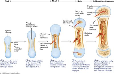
Endochondral ossification is the process by which most bones below the skull (except clavicles) are formed from a hyaline cartilage model. It involves primary and secondary ossification centers.
Bone collar forms around diaphysis of hyaline cartilage model.
Cartilage calcifies and develops cavities.
Periosteal bud invades cavities, forming spongy bone.
Diaphysis elongates; secondary ossification centers form in epiphyses.
Epiphyses ossify; hyaline cartilage remains at articular surfaces and epiphyseal plates.
Intramembranous Ossification
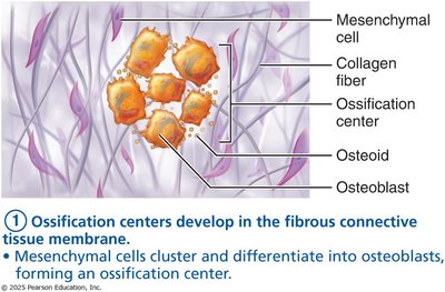
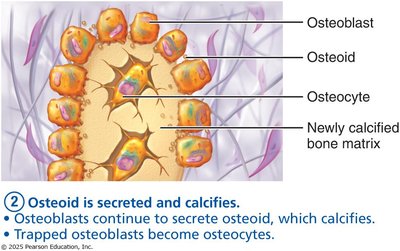
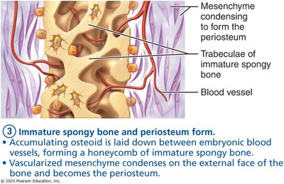
Intramembranous ossification forms flat bones, such as those of the skull and clavicles, from a fibrous membrane.
Ossification centers develop in fibrous connective tissue membrane.
Osteoid is secreted and calcifies; trapped osteoblasts become osteocytes.
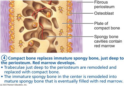
Immature spongy bone and periosteum form.
Compact bone replaces immature spongy bone; red marrow develops.
Bone Growth, Repair, and Remodeling
Postnatal Bone Growth
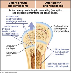
After birth, bones grow in length and width. Lengthwise growth occurs at the epiphyseal plate, where chondrocytes are replaced by bone. Growth in width (appositional growth) continues throughout adulthood, especially in response to mechanical stress.
Long Bone Growth and Remodeling
Bone growth and remodeling are balanced processes involving osteoblasts (bone deposition) and osteoclasts (bone resorption). Remodeling maintains bone shape and strength.
Regulation of Bone Growth
Hormones regulate bone growth:
Growth Hormone (GH): Stimulates epiphyseal plate changes during childhood.
Thyroid Hormone (TH): Ensures accurate bone proportions.
Sex Hormones (Estrogen and Testosterone): Promote growth spurts and epiphyseal plate closure during puberty.
Disorders of GH secretion can lead to gigantism (hypersecretion) or dwarfism (hyposecretion).
Bone Remodeling
Bone remodeling is a continuous process of bone resorption and deposition, ensuring bone health and adaptation to stress. About 5-10% of the skeleton is replaced yearly.
Skeletal System Homeostatic Mechanisms
Calcium Regulation
Bone acts as a calcium storage bank, and plasma calcium levels are regulated by hormones:
Parathyroid Hormone (PTH): Stimulates osteoclasts to release Ca2+ into the blood.
Calcitonin: Inhibits osteoclasts, promoting Ca2+ deposition in bone.
Calcitriol: Active form of vitamin D, increases calcium absorption from the gut.
Maintaining calcium homeostasis is essential for nerve function, muscle contraction, and blood clotting.
Mechanical Stress and Wolff’s Law
Wolff’s Law states that bone grows or remodels in response to the demands placed on it. Mechanical stress strengthens bone, while lack of movement leads to bone atrophy.
Abnormalities of the Skeletal System
Fractures
Fractures are classified by the position of bone ends (displaced vs. nondisplaced), completeness (complete vs. incomplete), and skin penetration (closed vs. open).
Reduction: Aligning bone ends (closed or open).
Repair: Involves hematoma formation, fibrocartilaginous callus formation, bony callus formation, and bone remodeling.
Osteomalacia and Rickets
Osteomalacia (adults) and rickets (children) are disorders characterized by poor bone mineralization, often due to vitamin D deficiency. Osteomalacia results in soft, weak bones, while rickets causes bone deformities and widened epiphyseal plates.
Osteoporosis
Osteoporosis is a condition where bone resorption exceeds bone deposition, leading to fragile, porous bones. It is common in postmenopausal women and increases the risk of fractures.
Risk Factors: Genetics, sex hormones, diet, insufficient bone stress, smoking, endocrine disorders, alcohol, medications.
Paget’s Disease
Paget’s disease involves abnormal bone remodeling, resulting in poorly mineralized, weak bones. It commonly affects adults over 40 and is often discovered incidentally on X-rays.
Symptoms: Pain, bone deformities, especially in the skull, spine, pelvis, and femur.
Causes: Unknown, possibly viral.
Additional info: The notes above expand on brief points from the original slides, providing definitions, examples, and academic context for each topic. All included images directly reinforce anatomical and physiological concepts discussed in the adjacent paragraphs.
