 Back
BackCardiovascular and Lymphatic Systems: Structure and Function
Study Guide - Smart Notes
 Tailored notes based on your materials, expanded with key definitions, examples, and context.
Cardiovascular and Lymphatic Systems
Introduction to Circulatory Systems
The human body relies on circulatory systems to transport essential materials such as oxygen, nutrients, and waste products. These systems are necessary due to the large number of cells and the size of the body, which make simple diffusion insufficient for material exchange. The circulatory system ensures that no cell is more than two cells away from a blood capillary, facilitating efficient transport and exchange.
Basic Components of Any Circulatory System
Four Fundamental Components
Vehicle: The substance that carries materials (e.g., blood or lymph).
Conduits: Channels or tubes (e.g., blood vessels, lymphatic vessels) through which the vehicle travels.
Motive Force: The pump or force that moves the vehicle (e.g., the heart).
Exchange Areas: Specialized regions where materials are exchanged (e.g., capillaries).
The Human Cardiovascular System
Major Components
Blood: The vehicle for oxygen, nutrients, and wastes.
Blood Vessels: The conduits for blood flow.
Heart: The muscular pump providing the motive force.
Capillaries: The exchange areas for gases, nutrients, and wastes.
Blood
Composition and Functions
Plasma: Makes up about 55% of blood volume; mainly water with dissolved proteins and other substances. After clotting, the remaining fluid is called serum.
Formed Elements: About 45% of blood volume; includes red blood cells (erythrocytes), white blood cells (leukocytes), and platelets (thrombocytes).
Red Blood Cells (Erythrocytes): Biconcave discs containing hemoglobin for oxygen transport; ~5 million/mm³ in adults.
White Blood Cells (Leukocytes): Immune cells; main types are neutrophils (phagocytosis) and lymphocytes (antibody production); ~5,000–11,000/mm³.
Platelets (Thrombocytes): Cell fragments involved in clotting; ~150,000–350,000/mm³.
Functions of Blood:
Transport of gases, nutrients, wastes, hormones, and heat
Temperature regulation
Immunity (via WBCs and antibodies)
Clotting to prevent blood loss and infection
Blood Vessels
General Structure and Types
Three-layered wall:
Intima: Inner smooth epithelial lining
Media: Middle layer of smooth muscle
Adventitia: Outer fibrous connective tissue
Types of Blood Vessels:
Arteries: Carry blood away from the heart
Veins: Carry blood toward the heart
Capillaries: Thin-walled vessels for exchange between blood and tissues
Valves: Present in veins and the heart to ensure one-way blood flow
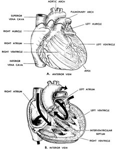
The Heart
Structure and Function
Location: Middle of the thorax, above the diaphragm, slightly to the left
Size: About the size of a clenched fist
Chambers: Four chambers—right and left atria (upper), right and left ventricles (lower)
Wall Layers:
Endocardium: Inner smooth epithelium
Myocardium: Middle cardiac muscle layer
Epicardium: Outer epithelium
Wall Thickness: Left ventricle is thickest due to higher pressure requirements
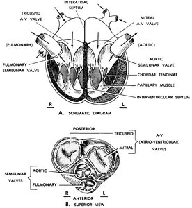
Cardiac Valves
Atrioventricular (A-V) Valves: Tricuspid (right), Mitral (left); prevent backflow into atria
Semilunar Valves: Pulmonary (right), Aortic (left); prevent backflow into ventricles
Chordae Tendineae and Papillary Muscles: Prevent valve prolapse
Control of Heartbeat
Extrinsic Nervous Control: Autonomic nerves (sympathetic accelerates, parasympathetic slows)
Intrinsic "Nervous" Control: Sinoatrial (S-A) node (pacemaker), Atrioventricular (A-V) node, septal bundles
Humoral Control: Blood-borne substances influence heart rate and strength
Coronary Circulation
Coronary Arteries: Supply the heart muscle with oxygenated blood
Cardiac Veins: Drain deoxygenated blood from the heart muscle into the right atrium
Clinical Note: Blockage of a coronary artery can cause tissue death (myocardial infarction)
Pericardium
Pericardial Sac: Serous membrane reducing friction during heartbeats
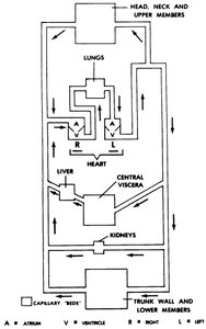
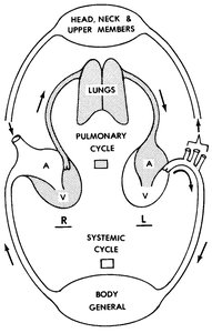
Cardiovascular Circulatory Patterns
Closed, Two-Cycle System
Closed System: Blood remains within vessels and heart
Pulmonary Cycle: Right heart → lungs → left heart (gas exchange)
Systemic Cycle: Left heart → body tissues → right heart (nutrient/waste exchange)
Collateral Circulation: Multiple vessels supply the same area for redundancy
End Artery: Single artery supplies a region; blockage leads to tissue death
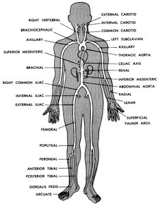
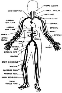
Major Arteries and Veins
Arteries: Carotid (head), subclavian (neck/upper limbs), aorta (trunk), iliac (pelvis/lower limbs)
Veins: Superior vena cava (head/upper limbs), inferior vena cava (rest of body), hepatic portal system (gut to liver)
Valves: Present in most veins, absent in veins from the head
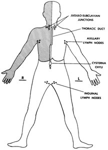
The Human Lymphatic System
General Function
The lymphatic system returns excess interstitial fluid (including proteins) to the bloodstream, maintaining fluid balance and contributing to immune defense.
Structures of the Lymphatic System
Lymphatic Capillaries: Absorb excess interstitial fluid in tissues
Lymph Vessels: Collect and transport lymph; contain valves to ensure one-way flow; major vessel is the thoracic duct
Lymph Nodes: Filter lymph and house immune cells
Tonsils: Lymphoid tissue at entrances to respiratory and digestive tracts for immune protection
Summary Table: Comparison of Cardiovascular and Lymphatic Systems
Feature | Cardiovascular System | Lymphatic System |
|---|---|---|
Vehicle | Blood | Lymph |
Conduits | Arteries, veins, capillaries | Lymphatic vessels, capillaries |
Motive Force | Heart | Skeletal muscle, vessel contraction |
Exchange Areas | Capillaries | Lymph nodes, capillaries |
Additional info: The lymphatic system also plays a key role in immune surveillance and fat absorption from the digestive tract (via lacteals).
