 Back
BackCardiovascular and Lymphatic Systems: Structure and Function Study Guide
Study Guide - Smart Notes
 Tailored notes based on your materials, expanded with key definitions, examples, and context.
Heart Anatomy
Structure of the Heart
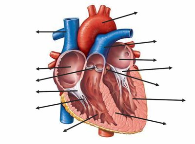
The heart is a muscular organ responsible for pumping blood throughout the body. It consists of four chambers: two atria (upper chambers) and two ventricles (lower chambers). Blood flows through a series of valves that ensure unidirectional movement and prevent backflow.
Right Atrium: Receives deoxygenated blood from the body via the vena cava.
Right Ventricle: Pumps blood to the lungs through the pulmonary artery.
Left Atrium: Receives oxygenated blood from the lungs via the pulmonary veins.
Left Ventricle: Pumps oxygenated blood to the body through the aorta.
Valves: Tricuspid, Pulmonary, Mitral (Bicuspid), and Aortic valves regulate blood flow between chambers and vessels.
Heart Physiology
Blood Flow Through the Heart
Blood follows a specific pathway through the heart, ensuring oxygenation and circulation:
Body → Vena Cava → Right Atrium → Tricuspid Valve → Right Ventricle → Pulmonary Valve → Lungs → Pulmonary Veins → Left Atrium → Mitral Valve → Left Ventricle → Aortic Valve → Aorta → Body
Right side: Pumps blood to the lungs (pulmonary circulation).
Left side: Pumps blood to the body (systemic circulation).
Intrinsic vs. Extrinsic Conduction System
The heart's rhythm is controlled by two systems:
Intrinsic (Internal): The heart generates its own electrical impulses via the SA node (pacemaker), AV node (delay), Bundle of His, Bundle Branches, and Purkinje fibers.
Extrinsic (External): The autonomic nervous system modulates heart rate. Sympathetic stimulation increases rate; parasympathetic (vagus nerve) decreases rate.
The Cardiac Cycle
The cardiac cycle describes the sequence of events in one heartbeat:
Ventricular Filling (Late Diastole): Heart relaxed, AV valves open, semilunar valves closed, blood flows into ventricles (80% filled).
Atrial Systole (Atrial Kick): Atria contract, push last 20% of blood into ventricles (End-Diastolic Volume, EDV).
Isovolumetric Contraction: Ventricles contract, AV valves close (S1, LUB), all valves closed, pressure rises, volume unchanged.
Ventricular Ejection: Semilunar valves open, blood leaves ventricles (End-Systolic Volume, ESV).
Isovolumetric Relaxation: Ventricles relax, semilunar valves close (S2, DUB), all valves closed, pressure drops.
Heart Sounds
S1 (LUB): Closing of AV valves at start of ventricular systole.
S2 (DUB): Closing of semilunar valves at start of ventricular diastole.
Heart sounds are caused by valve closure, not opening.
ECG and Cardiac Cycle
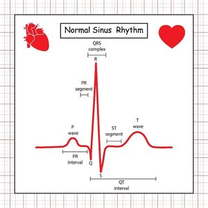
An electrocardiogram (ECG) records the electrical activity of the heart. Each wave corresponds to a specific event:
P wave: Atrial depolarization (contraction).
QRS complex: Ventricular depolarization (contraction).
T wave: Ventricular repolarization (relaxation).
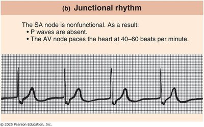
Junctional Rhythm: If the SA node fails, the AV node takes over, resulting in absent P waves and a slower heart rate (40-60 bpm).
Blood Vessels
Structure and Layers of Blood Vessels
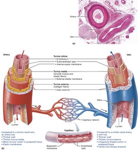
Blood vessels are classified by their structure and function. The wall consists of three layers:
Tunica Intima: Innermost layer, endothelium, smooth surface.
Tunica Media: Middle layer, smooth muscle and elastic fibers, controls diameter.
Tunica Externa (Adventitia): Outer layer, connective tissue, protection and anchoring.
Types of Blood Vessels
Arteries: Carry blood away from heart, thick tunica media, small lumen, high pressure, no valves.
Veins: Carry blood toward heart, thin walls, large lumen, low pressure, have valves.
Capillaries: Only tunica intima, site of exchange, very small.
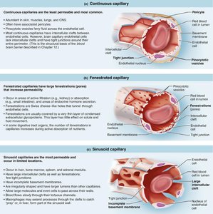
Types of Capillaries
Capillaries are specialized for exchange and differ in permeability:
Continuous Capillaries: Least permeable, tight junctions, found in skin, muscles, lungs, CNS.
Fenestrated Capillaries: Medium permeability, pores in endothelium, found in kidneys, small intestine, endocrine glands.
Sinusoid Capillaries: Most permeable, large gaps, found in liver, bone marrow, spleen, adrenal medulla.
Blood Flow, Pressure, and Resistance
Blood flow is determined by the relationship between pressure and resistance:
Formula:
Increasing pressure increases flow; increasing resistance decreases flow.
Total Peripheral Resistance (TPR): Resistance in systemic circulation, mainly controlled by arterioles.
Blood Pressure:
Mean Arterial Pressure (MAP)
MAP is the average pressure in arteries during one cardiac cycle:
Formula:
Where DP = Diastolic Pressure, SP = Systolic Pressure.
Blood Pressure Regulation and Baroreceptor Reflex
Blood pressure is regulated by cardiac output, vessel diameter, blood volume, viscosity, and arterial elasticity. The baroreceptor reflex provides short-term control:
Baroreceptors: Located in carotid sinus and aortic arch, detect changes in pressure.
High BP: Baroreceptors stretch, decrease sympathetic activity, heart rate slows, vessels dilate, BP decreases.
Low BP: Less stretch, increase sympathetic activity, heart rate increases, vessels constrict, BP increases.
Venous Return Mechanisms
Veins require assistance to return blood to the heart due to low pressure:
Valves: Prevent backflow, especially in legs.
Skeletal Muscle Pump: Muscle contractions squeeze veins, push blood upward.
Respiratory Pump: Inhalation decreases chest pressure, increases abdominal pressure, pulls blood toward heart.
Major Arteries and Veins
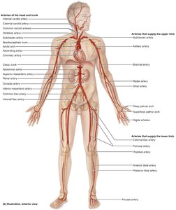
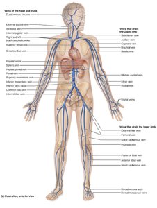
Major arteries and veins are responsible for distributing blood throughout the body. Arteries carry oxygenated blood away from the heart, while veins return deoxygenated blood.
Lymphatic System
Functions of the Lymphatic System
The lymphatic system maintains fluid balance, provides immune defense, and absorbs dietary fats:
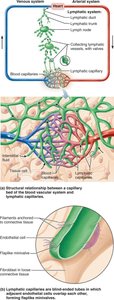
Returns fluid: Picks up excess interstitial fluid and returns it to the bloodstream.
Immune defense: Filters lymph, removes pathogens, houses lymphocytes.
Absorbs fats: Lacteals in the small intestine absorb dietary fats.
Lymphoid Cells: T and B Lymphocytes
B Lymphocytes (B Cells): Mature in bone marrow, produce antibodies, fight pathogens in body fluids.
T Lymphocytes (T Cells): Mature in thymus, attack infected or abnormal cells directly.
Both originate: In red bone marrow.
Lymphoid Tissues and Organs
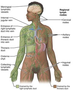
Lymphoid tissues: Contain lymphocytes and immune cells, monitor body fluids, fight infection.
Lymph nodes: Small, bean-shaped organs that filter lymph and activate immune cells.
Primary lymphoid organs: Red bone marrow and thymus (site of lymphocyte formation and maturation).
Secondary lymphoid organs: Lymph nodes, spleen, tonsils, MALT (site of immune activation).
Lymph Node Structure and Circulation
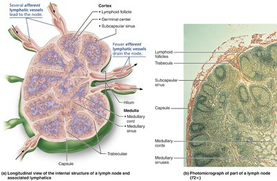
Lymph flows through nodes in a specific sequence:
Afferent lymphatic vessels bring lymph into the node.
Lymph enters the subcapsular sinus.
Flows through cortex (B cells).
Flows through medulla (macrophages clean it).
Efferent lymphatic vessel exits the node.
Spleen and MALT
Spleen: Removes old red blood cells, activates immune response, stores platelets and blood.
MALT: Mucosa-Associated Lymphoid Tissue, protects mucous membranes, includes Peyer's patches in the small intestine.
Thymus
Location: Superior mediastinum, behind sternum, above heart.
Function: Site of T cell maturation, teaches self vs. non-self, prevents autoimmunity.
Large in children, shrinks after puberty, does not filter lymph.
