Back
Cardiovascular Physiology: Structure, Function, and Regulation
Study Guide - Smart Notes
 Tailored notes based on your materials, expanded with key definitions, examples, and context.
Cardiovascular System Overview
Structure and Function
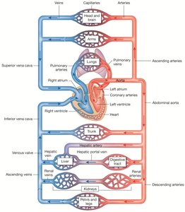
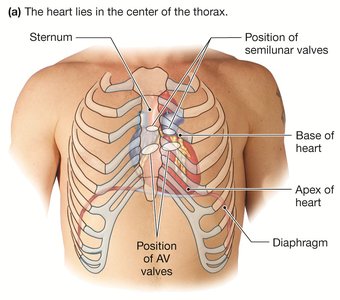
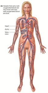
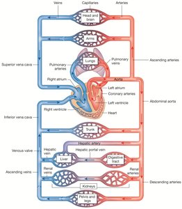
The cardiovascular system is a closed system responsible for transporting blood throughout the body. It consists of the heart (the pump), blood (the fluid), and blood vessels (the tubes). The heart has four chambers: two atria and two ventricles.
Heart: Central pump that maintains blood flow.
Blood Vessels: Arteries, veins, and capillaries that carry blood to and from tissues.
Blood: Transports nutrients, gases, hormones, immune cells, and waste products.
Transport Functions
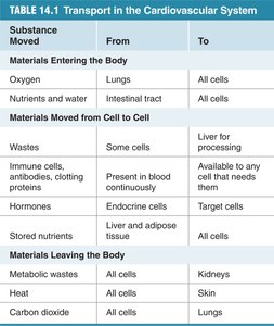
The cardiovascular system facilitates the movement of materials between the external environment and cells, as well as between cells.
Materials Entering the Body: Oxygen (lungs to all cells), nutrients and water (intestinal tract to all cells).
Materials Moved from Cell to Cell: Wastes, immune cells, antibodies, hormones, stored nutrients.
Materials Leaving the Body: Metabolic wastes (to kidneys), heat (to skin), carbon dioxide (to lungs).
Substance Moved | From | To |
|---|---|---|
Oxygen | Lungs | All cells |
Nutrients and water | Intestinal tract | All cells |
Wastes | Some cells | Liver for processing |
Immune cells, antibodies, clotting proteins | Present in blood continuously | Available to any cell that needs them |
Hormones | Endocrine cells | Target cells |
Stored nutrients | Liver and adipose tissue | All cells |
Metabolic wastes | All cells | Kidneys |
Heat | All cells | Skin |
Carbon dioxide | All cells | Lungs |
Circulatory Pathways
Systemic Circulation
Systemic circulation carries oxygenated blood from the left ventricle through the aorta to the body's tissues and organs, and returns deoxygenated blood to the right atrium via systemic veins.
Arteries: Carry oxygen-rich blood from the heart to tissues.
Veins: Return deoxygenated blood to the heart.
Pulmonary Circulation
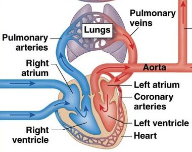
Pulmonary circulation transports deoxygenated blood from the right ventricle to the lungs via pulmonary arteries, and returns oxygenated blood to the left atrium via pulmonary veins.
Pulmonary Arteries: Carry blood from the right ventricle to the lungs.
Pulmonary Veins: Return oxygen-rich blood to the left atrium.
Embryonic Heart Development
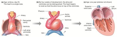
The heart develops from a single tube in the embryo, which later differentiates into chambers and valves.
Early Development: Single tube structure.
Chamber Formation: Atria and ventricles become distinguishable by four weeks.
Cardiac Output and Blood Flow
Cardiac Output (Q̇)
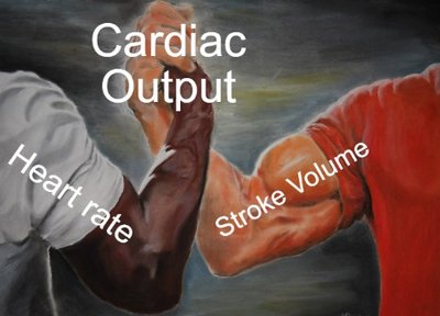
Cardiac output is the amount of blood pumped by the heart per minute. It is a key indicator of the heart's ability to meet the body's demand for blood flow.
Formula:
At Rest: Approximately 5 L/min.
Pressure, Flow, and Resistance
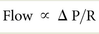
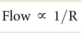
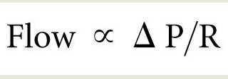
Blood flow is governed by pressure gradients and resistance, following Ohm's Law.
Flow Equation:
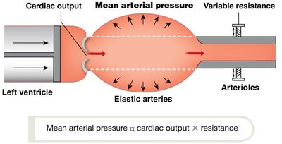
Physiological Equivalent:
MAP: Mean Arterial Pressure (driving force for blood flow).
TPR: Total Peripheral Resistance (determined by vessel diameter).
Blood Pressure Gradient
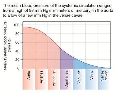
Blood flows down a pressure gradient, from high pressure in the arteries to low pressure in the veins.
Highest Pressure: Aorta (93 mm Hg).
Lowest Pressure: Venae cavae (few mm Hg).
Exercise, Hypoxia, and Oxygen Transport
Exercise and Hypoxia
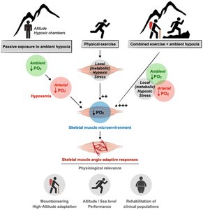
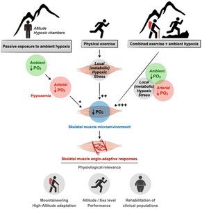
During exercise, especially at high altitude, oxygen transport is challenged by reduced partial pressure of oxygen (PO2).
Oxygen Gradient: Moves from high PO2 in arterial blood to low PO2 in tissues.
Hypoxia: Reduces efficiency of oxygen delivery, limiting aerobic metabolism.
Cellular Adaptations to Hypoxia
Hypoxia induces changes in glucose transporters and upregulates genes involved in oxygen transport and mitochondrial efficiency.
HIF-1-alpha: Upregulates oxygen transport genes.
PGC-1α: Increases mitochondrial content and efficiency.
Regulation of Blood Flow and Resistance
Resistance in Blood Vessels
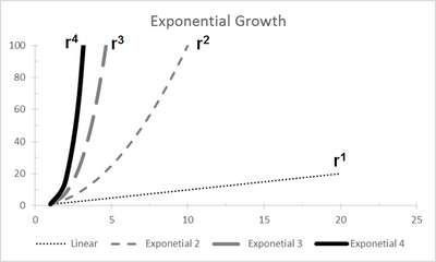
Resistance opposes blood flow and is influenced by vessel length, radius, and blood viscosity.
Length (L): Resistance increases with vessel length.
Radius (r): Resistance decreases exponentially as radius increases ().
Viscosity (η): Resistance increases with higher viscosity.
Vasoconstriction and Vasodilation
Blood vessel radius is physiologically regulated through vasoconstriction and vasodilation.
Vasoconstriction: Decreases radius, increases resistance, decreases blood flow.
Vasodilation: Increases radius, decreases resistance, increases blood flow.
Example: Beetroot juice enhances vasodilation via nitric oxide pathway, improving oxygen delivery to muscles.
Summary Table: Key Equations and Concepts
Cardiac Output:
Flow:
Mean Arterial Pressure:
Resistance:
Conclusion
The cardiovascular system is essential for maintaining homeostasis, delivering oxygen and nutrients, and removing waste products. Its function is tightly regulated by pressure gradients, resistance, and physiological adaptations to exercise and hypoxia.