 Back
BackCardiovascular System: Blood, Heart, and Circulation – Study Notes
Study Guide - Smart Notes
 Tailored notes based on your materials, expanded with key definitions, examples, and context.
Blood: Composition and Function
Importance and Composition of Blood
Blood is a vital connective tissue responsible for transporting nutrients, gases, hormones, and waste products throughout the body. It consists of two main components: formed elements (about 45%) and plasma (about 55%).
Plasma: The nonliving fluid matrix, primarily water (90%), with dissolved nutrients, gases, hormones, waste, proteins, and electrolytes. Its composition varies as cells exchange substances with the blood.
Formed Elements: Includes erythrocytes (red blood cells), leukocytes (white blood cells), and platelets.
Clotting: Blood fibers become visible during clotting, a key property of connective tissue.
Types of Blood Cells
Erythrocytes (Red Blood Cells): Sacs of hemoglobin that transport oxygen and a small amount of carbon dioxide.
Leukocytes (White Blood Cells): Defend the body against infection and are part of the immune system.
Platelets: Cell fragments involved in hemostasis (blood clot formation).
Hematologic Tests
Total White Blood Cell Count: Measures WBCs per unit volume. Leukocytosis (high WBC) may indicate infection; Leukopenia (low WBC) may result from toxins; Leukemia is uncontrolled WBC proliferation.
Total Red Blood Cell Count: Assesses oxygen transport capacity; low counts may indicate anemia.
Blood Typing: Based on genetically determined glycoproteins and antibodies present in plasma.
Cholesterol Concentration: High levels can lead to atherosclerosis, where blood vessels are blocked by plaques.
The Cardiovascular System
General Function
The cardiovascular system transports oxygen, nutrients, wastes, and other substances via blood, propelled by the heart's contractions.
Anatomy of the Human Heart
Pericardium: Double-walled sac enclosing the heart; serous fluid reduces friction.
Heart Wall Layers:
Epicardium: Outer layer (visceral pericardium).
Myocardium: Middle, muscular layer reinforced by connective tissue.
Endocardium: Inner lining, continuous with blood vessels and covering valves.
Heart Chambers and Valves
Chambers: 2 atria (receiving), 2 ventricles (discharging).
Septum: Interatrial and interventricular septa divide the heart longitudinally.
Valves: Four valves ensure one-way blood flow:
Atrioventricular (AV) Valves: Tricuspid (right), Mitral (left).
Semilunar Valves: Pulmonary and aortic valves.
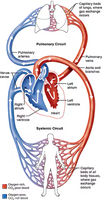
Pulmonary, Systemic, and Coronary Circulations
The heart acts as a double pump, supporting three main circulatory routes:
Pulmonary Circuit: Right heart pumps oxygen-poor blood to the lungs for gas exchange.
Systemic Circuit: Left heart pumps oxygen-rich blood to the body.
Coronary Circulation: Coronary arteries supply the heart muscle itself.
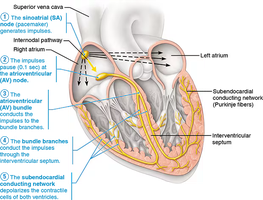
Conduction System of the Heart
Heart contractions are coordinated by an intrinsic conduction system, allowing the heart to function as a unit.
Sinoatrial (SA) Node: Pacemaker, initiates impulses.
Atrioventricular (AV) Node: Delays impulse, allowing atrial contraction.
AV Bundle (Bundle of His): Conducts impulses to bundle branches.
Bundle Branches: Carry impulses through interventricular septum.
Subendocardial Conducting Network (Purkinje fibers): Depolarizes ventricular muscle cells.
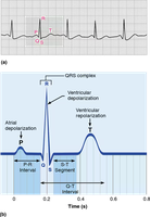
Electrocardiography (ECG/EKG)
The heart's electrical activity is recorded as an electrocardiogram, which displays three main waves:
P wave: Atrial depolarization.
QRS Complex: Ventricular depolarization.
T wave: Ventricular repolarization.
Segments: Regions between waves.
Intervals: Regions containing segments and one or more waves.
Blood Vessels and Circulation
Structure of Blood Vessels
Arteries: Carry blood away from the heart; have thicker walls to withstand higher pressure.
Veins: Return blood to the heart; have thinner walls and often valves to prevent backflow.
Three Tunics (Layers):
Tunica Intima: Endothelial lining, reduces resistance.
Tunica Media: Smooth muscle, regulates vessel diameter and blood pressure.
Tunica Externa: Connective tissue, provides support and protection.
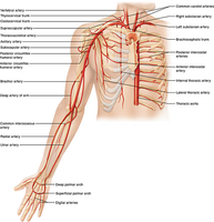
Major Systemic Arteries
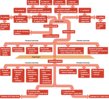
Systemic arteries are generally deep and well-protected. The aorta is the largest artery, branching into the ascending aorta, aortic arch, and descending aorta.
Major Systemic Veins
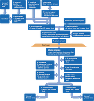
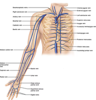
Systemic veins are more superficial and converge on the superior and inferior venae cavae. Veins draining the head and upper extremities empty into the superior vena cava, while those from the lower body empty into the inferior vena cava.
Special Circulations
Pulmonary Circulation: Brings blood into close contact with alveoli for gas exchange; does not serve tissue metabolic needs.
Fetal Circulation: The fetus exchanges gases and nutrients via the placenta, as the lungs and digestive system are nonfunctional.
Hepatic Portal Circulation: Blood from digestive organs is routed to the liver via the hepatic portal vein for processing before entering systemic circulation.
Cardiac Cycle and Heart Function
Cardiac Cycle
Systole: Ventricular contraction phase.
Diastole: Ventricular relaxation phase.
Cardiac Cycle: One complete heartbeat, including both systole and diastole.
Quiescent Period: Total heart relaxation.
Heart Sounds
Produced by turbulent blood flow as valves close during the cardiac cycle.
Abnormal sounds (murmurs) may indicate valve or circulatory issues.
The Pulse
Pulse: Alternating surges of pressure in an artery (expansion and recoil).
Pulse Pressure: Difference between systolic and diastolic pressure.
Apical Pulse: Felt at the heart's apex; may be slightly faster than the radial pulse.
Radial Pulse: Felt at the radial artery.
Pulse Deficit: Difference between apical and radial pulses.
Blood Pressure
Blood Pressure: Force exerted by blood against vessel walls.
Systolic Pressure: Pressure during ventricular contraction.
Diastolic Pressure: Pressure during ventricular relaxation.
Formula:
Skin Color as an Indicator of Circulatory Dynamics
Skin color reflects local circulation and can indicate systemic circulatory status.
Pale, cold, and moist skin may suggest diverted blood flow or circulatory compromise.
