 Back
BackCardiovascular System: Circulation Pathways and Blood Pressure Regulation
Study Guide - Smart Notes
 Tailored notes based on your materials, expanded with key definitions, examples, and context.
Blood Vessels: Structure and Function
Types of Blood Vessels
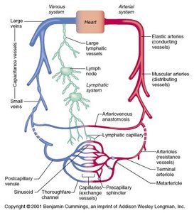
The cardiovascular system is composed of three main types of blood vessels: arteries, veins, and capillaries. Each type has a distinct structure and function in the circulation of blood throughout the body.
Arteries: Conduct blood away from the heart. Subtypes include elastic arteries, muscular arteries, and arterioles.
Veins: Conduct blood toward the heart. Subtypes include venules, small veins, and large veins.
Capillaries: The thinnest blood vessels, serving as the primary site for exchange between blood and tissues. Capillaries form networks called capillary beds and are the functional units of the circulatory system (microcirculation).
Capillary Anatomy and Types
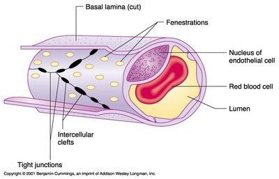
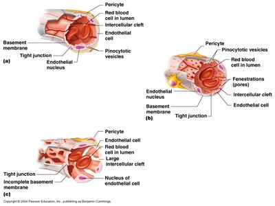
Capillaries are specialized for exchange and have three main structural types, each adapted for specific functions:
Continuous (Tight) Capillaries: Have uninterrupted endothelial lining with tight junctions; found in muscle, skin, and the brain.
Fenestrated Capillaries: Possess pores (fenestrations) that increase permeability; common in kidneys, intestines, and endocrine glands.
Sinusoidal Capillaries: Have large gaps and an incomplete basement membrane, allowing passage of large molecules and cells; found in the liver, bone marrow, and spleen.
Capillary Beds and Microcirculation
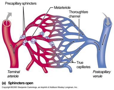
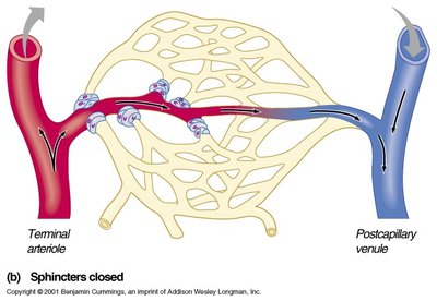
Capillary beds are networks of capillaries supplied by arterioles and drained by venules. Blood flow through these beds is regulated by precapillary sphincters and can be shunted via metarterioles and thoroughfare channels.
Precapillary Sphincters: Rings of smooth muscle that regulate blood flow into true capillaries.
Metarterioles: Short vessels linking arterioles and capillaries, providing a direct route for blood flow when sphincters are closed.
Capillary Transport Pathways
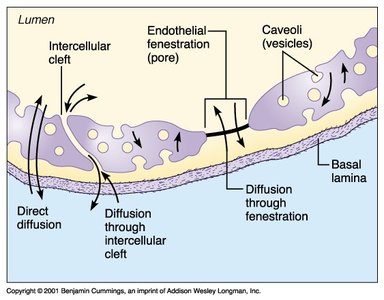
Substances move across capillary walls by several mechanisms:
Direct Diffusion: Lipid-soluble substances pass directly through endothelial cell membranes.
Intercellular Clefts: Water-soluble substances pass through gaps between endothelial cells.
Fenestrations: Pores allow rapid passage of small molecules.
Vesicular Transport: Large molecules are transported via vesicles (caveolae).
Forces Affecting Bulk Flow Across Capillary Walls
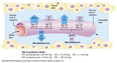
Bulk flow across capillary walls is governed by hydrostatic and osmotic pressures, collectively known as Starling forces. These determine the movement of fluid into and out of capillaries.
Hydrostatic Pressure (HP): The force exerted by fluid pressing against a wall; pushes fluid out of capillaries.
Osmotic Pressure (OP): The force exerted by proteins drawing water into the capillaries.
Net Filtration Pressure (NFP): The balance of these forces determines whether fluid leaves or enters the capillary.
Equation:
Where: = capillary hydrostatic pressure = interstitial fluid osmotic pressure = interstitial fluid hydrostatic pressure = capillary osmotic pressure
Arteries and Veins: Structure and Function
Comparative Anatomy of Arteries and Veins
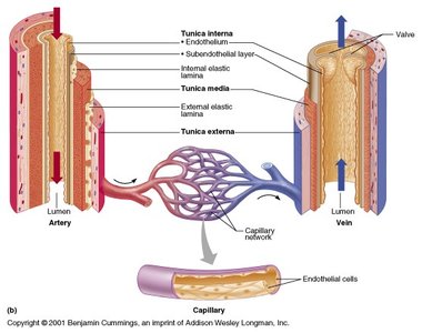
Arteries and veins have distinct structural differences that reflect their functions in the circulatory system.
Arteries: Known as resistance vessels, especially arterioles. They have thick walls to withstand high pressure and regulate blood flow.
Veins: Known as capacitance vessels, they have thinner walls and larger lumens, allowing them to hold more blood and adjust volume as needed.
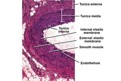
Summary Table: Blood Vessel Anatomy
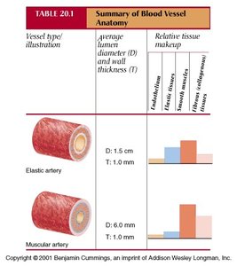
The following tables summarize the structural differences among major blood vessel types:
Vessel Type | Average Lumen Diameter (D) | Wall Thickness (T) | Relative Tissue Makeup |
|---|---|---|---|
Elastic artery | 1.5 cm | 1.0 mm | High elastic tissue, moderate smooth muscle, low fibrous tissue |
Muscular artery | 6.0 mm | 1.0 mm | High smooth muscle, moderate elastic tissue, low fibrous tissue |
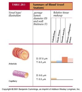
Vessel Type | Average Lumen Diameter (D) | Wall Thickness (T) | Relative Tissue Makeup |
|---|---|---|---|
Arteriole | 37.0 μm | 6.0 μm | Moderate smooth muscle, low elastic and fibrous tissue |
Capillary | 9.0 μm | 0.5 μm | Endothelium only |
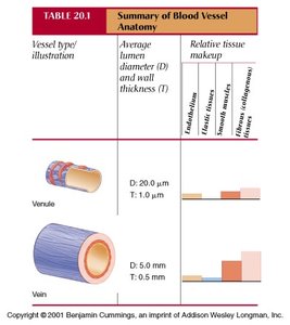
Vessel Type | Average Lumen Diameter (D) | Wall Thickness (T) | Relative Tissue Makeup |
|---|---|---|---|
Venule | 20.0 μm | 1.0 μm | Low smooth muscle, low elastic tissue, moderate fibrous tissue |
Vein | 5.0 mm | 0.5 mm | Low smooth muscle, low elastic tissue, high fibrous tissue |
Hemodynamics: Blood Flow, Pressure, and Resistance
Blood Pressure Through the Vascular Tree
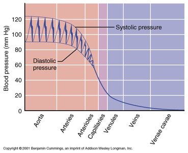
Blood pressure (BP) decreases as blood moves from arteries to veins. The greatest drop occurs in the arterioles, which are the main resistance vessels.
Systolic Pressure (SP): Pressure during ventricular contraction.
Diastolic Pressure (DP): Pressure during ventricular relaxation.
Mean Arterial Pressure (MAP): Weighted average pressure in the arteries.
Equations:
Where: = Cardiac Output = Total Peripheral Resistance
Total Peripheral Resistance (TPR)
TPR is the opposition to blood flow due to friction within blood vessels. Arteriolar diameter is the most significant factor affecting TPR, regulated by sympathetic innervation and vessel compliance. Blood viscosity also contributes to resistance.
Blood Flow Velocity in the Vascular Tree
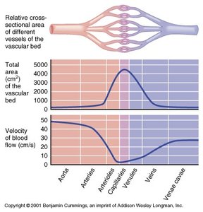
Blood flow velocity is inversely related to the total cross-sectional area of the vessels. It is slowest in the capillaries, allowing for efficient exchange of materials.
Blood Flow Changes During Exercise
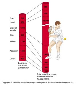
During exercise, blood flow is redistributed to meet the increased metabolic demands of skeletal muscle, heart, and skin, while flow to other organs may decrease.
Clinical Correlations
Laminar vs. Turbulent Blood Flow
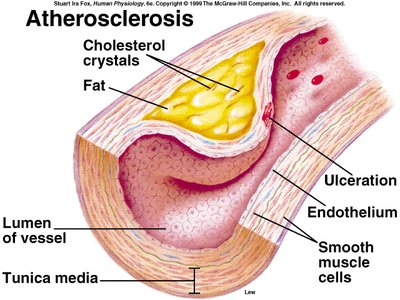
In healthy vessels, blood flow is typically laminar (smooth and silent). In diseased vessels, such as those with atherosclerotic plaques, flow becomes turbulent (rough and noisy), which can be detected as abnormal sounds during blood pressure measurement.
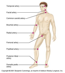
Pressure Points
Certain superficial arteries can be compressed to control bleeding or to measure pulse. These are known as pressure points.
Venous Return and Regulation
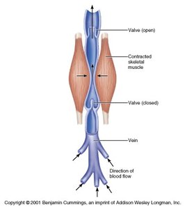
Skeletal Muscle Pump
Venous return to the heart is aided by the skeletal muscle pump, which uses muscle contractions to propel blood toward the heart, assisted by one-way valves in veins.
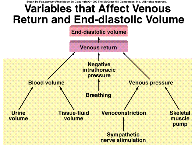
Factors Affecting Cardiac Output (CO)
Cardiac output is influenced by heart rate, stroke volume, venous return, and autonomic nervous system activity. These factors are integrated to maintain adequate tissue perfusion and blood pressure.
Summary of Key Equations
Blood Pressure:
Pulse Pressure:
Mean Arterial Pressure:
MAP and Cardiac Output:
Renal and Hormonal Control of Blood Pressure
The kidneys and various hormones (such as the renin-angiotensin-aldosterone system) play crucial roles in long-term regulation of blood pressure by adjusting blood volume and vessel tone.
Summary Table: Factors Affecting MAP
Factor | Effect on MAP |
|---|---|
Cardiac Output (CO) | Directly proportional |
Total Peripheral Resistance (TPR) | Directly proportional |
Blood Volume | Directly proportional |
Vessel Diameter | Inversely proportional to resistance |
Additional info: This guide integrates and expands upon the provided lecture content, including definitions, mechanisms, and clinical relevance for ANP college students.
