 Back
BackCell Structure and Function: Comprehensive Study Notes for ANP College Students
Study Guide - Smart Notes
 Tailored notes based on your materials, expanded with key definitions, examples, and context.
Cell Structure and Function
Introduction to Cells and Cell Theory
Cells are the fundamental units of life, forming the basis of all living organisms. The cell theory outlines the essential properties of cells, which are crucial for understanding anatomy and physiology.
Cell Theory:
Cells are the building blocks of plants and animals.
Cells are the smallest functioning units of life.
Cells are produced through division of preexisting cells.
Each cell maintains homeostasis.
Cytology: The study of cell structure and function.
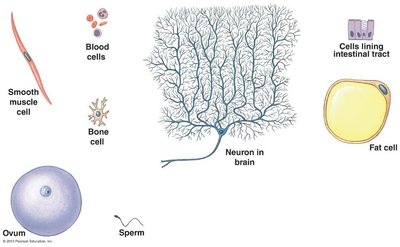
Cell Diversity: Human cells vary greatly in structure and function, each specialized for particular roles.
Overview of Cell Anatomy
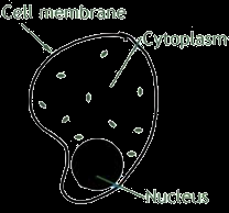
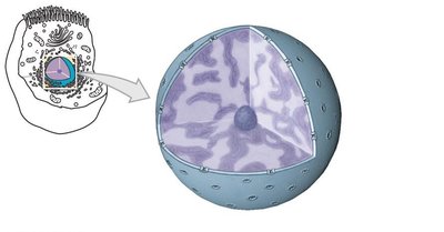
Cells are enclosed by a plasma membrane and contain cytoplasm, which houses various organelles. The cytoplasm consists of cytosol (fluid) and organelles (structures with specific functions).
Plasma Membrane: Surrounds every cell, composed of lipids and proteins, provides protection, and regulates movement of molecules.
Cytoplasm: Includes cytosol and organelles.
Cytosol: The intracellular fluid containing ions, proteins, carbohydrates, amino acids, lipids, and inclusions (e.g., glycogen, lipid droplets).
Intracellular Fluid (ICF): Fluid inside the cell.
Extracellular Fluid (ECF): Fluid outside the cell, including interstitial fluid and plasma.

Cell Organelles
Nucleus
The nucleus is the control center of the cell, storing and protecting DNA. It is typically the largest organelle and contains several structures.
Nuclear Envelope: Double membrane surrounding the nucleus.
Nuclear Pores: Allow exchange of materials between nucleus and cytoplasm.
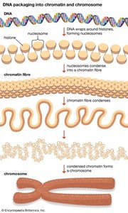
DNA: Contains genetic code for protein synthesis, organized into 23 pairs of chromosomes in humans.
Nucleoplasm: Gel-like fluid inside the nucleus.
Nucleolus: Produces ribosomal RNA (rRNA).
Chromatin: Loosely coiled DNA and proteins when the cell is not dividing.
Ribosomes
Ribosomes are non-membranous organelles responsible for protein synthesis. They can be attached to the rough endoplasmic reticulum (RER) or free in the cytoplasm.
Structure: Composed of two subunits (large and small), made of rRNA and protein.
Function: Bind mRNA and tRNAs, catalyze peptide bond formation.
Large Subunit (LSU): Contains peptidyl transferase center.
Small Subunit (SLU): Binds mRNA and decodes genetic information.
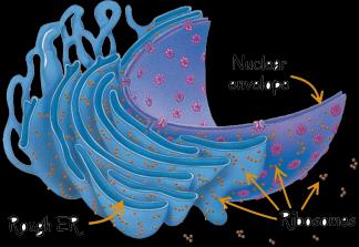
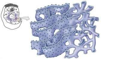
Endoplasmic Reticulum (ER)
The ER is a network of membranous sheets and channels, continuous with the nuclear envelope. It is divided into two types: smooth and rough.
Smooth ER (SER): Lacks ribosomes, involved in lipid synthesis, hormone production, detoxification, and carbohydrate metabolism.
Rough ER (RER): Studded with ribosomes, processes and modifies proteins for secretion or membrane insertion.
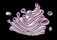
Golgi Apparatus
The Golgi apparatus is a stack of flat membrane sacs that functions as the cell's 'post office,' collecting, sorting, packaging, and distributing proteins and lipids.
Function: Modifies and packages proteins from the RER, directs them to their destinations.
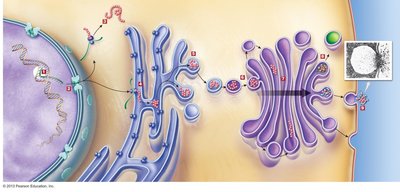
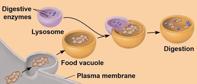
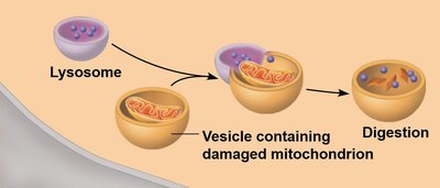
Lysosomes
Lysosomes are vesicles produced by the Golgi body, containing hydrolytic enzymes for digestion.
Function: Break down ingested food, damaged organelles, and foreign particles.
Peroxisomes
Peroxisomes are specialized membrane-bound organelles involved in catabolism of fatty acids and detoxification.
Function: Generate and degrade hydrogen peroxide, neutralized by catalase enzyme.

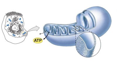
Mitochondria
Mitochondria are rod-shaped organelles with a double membrane and internal folds called cristae. They are the site of cellular respiration and ATP synthesis.
Structure: Outer membrane, inner membrane with cristae, matrix.
Function: Produce ATP, house enzymes for respiration.
Cytoskeleton
The cytoskeleton is a network of filaments and tubules that organizes cell structure and activities.
Components: Microtubules, microfilaments, intermediate filaments.
Functions: Maintain cell shape, anchor organelles, assist in movement.
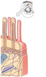
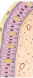
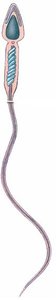
Cilia and Flagella
Cilia and flagella are extensions of the cell surface made of microtubules. Cilia move fluids over the cell surface, while flagella enable cell movement.
Cilia: Short, numerous, move mucus and dust in respiratory passages.
Flagella: Long, usually one per cell (e.g., sperm), enable cell movement.
Microvilli
Microvilli are finger-shaped projections formed by folding of the plasma membrane, increasing surface area for absorption.
Location: On cells actively absorbing substances.
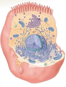
Centrioles and Centrosome
Centrosomes are organelles made of two centrioles at right angles, essential for chromosome movement during cell division.
Function: Organize microtubule network, initiate cell division processes.

Plasma Membrane and Transport Mechanisms
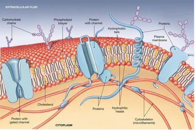
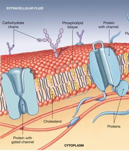
Plasma Membrane Structure
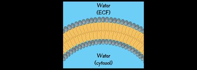
The plasma membrane is a strong, flexible barrier composed of lipids and proteins, described by the Fluid Mosaic model.
Phospholipid Bilayer: Two layers with hydrophilic heads facing water and hydrophobic tails facing inward.
Cholesterol: Embedded in the bilayer, maintains fluidity and prevents close packing at low temperatures.
Proteins:
Transmembrane proteins span the membrane.
Peripheral proteins are partially embedded on either side.
Carbohydrates: Attached to proteins (glycoproteins) or lipids (glycolipids), involved in cell-cell identification.


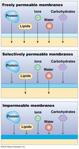
Membrane Permeability
Permeability refers to the ease with which substances cross the plasma membrane. It can be freely permeable, selectively permeable, or impermeable.
Factors Affecting Permeability:
Size and shape of molecule
Electric charge
Lipid solubility
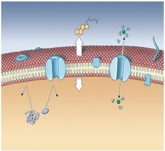
Membrane Transport Mechanisms
Transport across the membrane can be passive (no energy required) or active (requires ATP).
Passive Transport: Moves substances down their concentration gradient (e.g., diffusion, osmosis, facilitated diffusion).
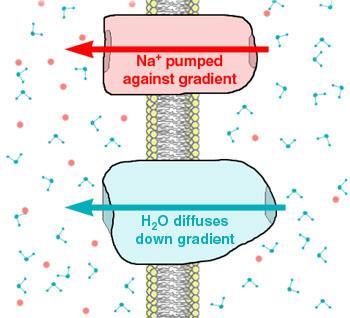
Active Transport: Moves substances against their concentration gradient, requires ATP (e.g., solute pumping, vesicular transport).
Diffusion
Diffusion is the movement of solute molecules from higher to lower concentration, important for eliminating concentration gradients in the body.
Lipid-soluble molecules: Diffuse easily through the membrane.
Ions and water-soluble substances: Require membrane channels.
Example: Transport of oxygen and CO2 between lungs and blood.
Osmosis
Osmosis is the diffusion of water across a selectively permeable membrane, from higher to lower water concentration.
Osmotic Pressure: The force with which water moves into a solution due to solute concentration.
Tonicity:
Hypertonic: Higher solute concentration outside cell, cell shrivels (crenation).
Hypotonic: Lower solute concentration outside cell, cell swells and may lyse (hemolysis).
Isotonic: Equal solute concentration, no net movement.
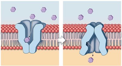
Carrier-Mediated Transport
Carrier-mediated transport uses specialized membrane proteins to move substrates and ions, either passively (facilitated diffusion) or actively (active transport).
Facilitated Diffusion: Carrier proteins move larger molecules (e.g., glucose) down their concentration gradient without ATP.
Active Transport: Uses ATP to move substances against their gradient, often via pumps (e.g., sodium-potassium pump).
Exchange Pump: Moves two ions in opposite directions, e.g., 3 Na+ out and 2 K+ in per ATP used.
Vesicular Transport
Vesicular transport moves substances in membrane sacs, requiring energy. Includes endocytosis (import) and exocytosis (export).
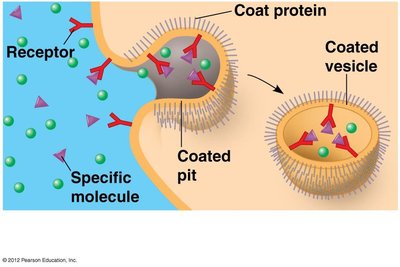
Endocytosis:
Receptor-mediated: Molecules bind to receptors, membrane pinches off to form vesicle.
Pinocytosis: 'Cell drinking,' vesicles filled with fluid.
Phagocytosis: 'Cell eating,' vesicles with large particles.
Exocytosis: Vesicles fuse with plasma membrane, releasing contents outside the cell (e.g., insulin secretion).
Summary Table: Membrane Transport Types
Transport Type | Energy Required | Direction | Example |
|---|---|---|---|
Diffusion | No | Down gradient | O2/CO2 exchange |
Osmosis | No | Down water gradient | Water movement |
Facilitated Diffusion | No | Down gradient | Glucose transport |
Active Transport | Yes (ATP) | Against gradient | Na+/K+ pump |
Endocytosis | Yes (ATP) | Into cell | Receptor-mediated |
Exocytosis | Yes (ATP) | Out of cell | Insulin secretion |
Key Equations
Osmotic Pressure: Where is osmotic pressure, is the van't Hoff factor, is molarity, is the gas constant, and is temperature.
Diffusion Rate (Fick's Law): Where is flux, is diffusion coefficient, is concentration gradient.
Conclusion
Understanding cell structure and function is fundamental to anatomy and physiology. The plasma membrane, organelles, and transport mechanisms work together to maintain cellular homeostasis and support life processes.
