 Back
BackCell Structure, Organelles, and Membrane Transport: ANP College Study Guide
Study Guide - Smart Notes
 Tailored notes based on your materials, expanded with key definitions, examples, and context.
Cell Structure and Function
Overview of Cell Anatomy
Cells are the fundamental units of life, each enclosed by a plasma membrane and containing cytoplasm. The cytoplasm consists of cytosol (the fluid part) and organelles (specialized structures performing distinct functions). The study of cell structure and function is called cytology. - Plasma membrane: Surrounds the cell, providing protection and controlling molecular movement. - Cytoplasm: Includes cytosol and organelles. - Nucleus: The control center of the cell, storing genetic material. 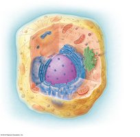
Diversity of Cell Types
Cells vary greatly in structure and function, adapted to their specific roles in the body. Examples include muscle cells for movement, nerve cells for communication, and blood cells for transport. 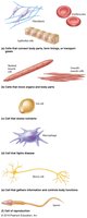
Cell Organelles
Classification of Organelles
Organelles are classified based on the presence or absence of a membrane: - Membranous organelles: Surrounded by a membrane, compartmentalizing functions (e.g., nucleus, mitochondria, lysosomes, endoplasmic reticulum, Golgi apparatus, peroxisomes). - Non-membranous organelles: Not surrounded by a membrane; components are in direct contact with cytoplasm (e.g., ribosomes, cytoskeleton, cilia).
Nucleus
The nucleus is typically the largest organelle, containing: - Nuclear envelope: Double membrane with nuclear pores. - DNA: Genetic material organized in chromosomes (23 pairs in humans). - Nucleoplasm: Fluid inside the nucleus. - Nucleolus: Site of ribosomal RNA synthesis. 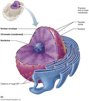
Function of the Nucleus
- Stores and protects DNA. - Controls cell function by regulating protein synthesis. - DNA is tightly coiled in chromosomes during cell division; otherwise, it exists as chromatin. - The nucleolus produces ribosomal RNA for ribosome assembly. 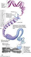
Ribosomes
Ribosomes are non-membranous organelles composed of two subunits (large and small), made of RNA and protein. - Function: Protein synthesis. - Location: Attached to rough ER or free in cytoplasm. 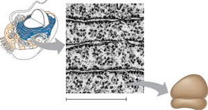
Endoplasmic Reticulum (ER)
The ER is a network of membranous sheets and channels, continuous with the nuclear envelope. - Smooth ER (SER): Lacks ribosomes; synthesizes and breaks down fats and carbohydrates, synthesizes steroids, detoxifies toxins. - Rough ER (RER): Studded with ribosomes; processes and modifies proteins, forms membranes. 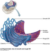
Golgi Apparatus
The Golgi apparatus is a stack of flat membrane sacs. - Function: Collects, sorts, packages, and distributes proteins and lipids. - Works with ribosomes, RER, and vesicles to process and transport proteins. 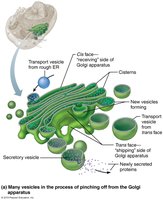
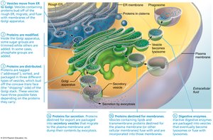
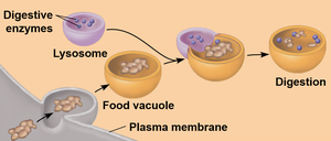
Lysosomes
Lysosomes are vesicles produced by the Golgi body, containing hydrolytic enzymes. - Function: Digest ingested food, damaged organelles, and foreign particles. 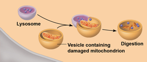
Mitochondria
Mitochondria are rod-shaped organelles with a double membrane. - Outer membrane: Smooth. - Inner membrane: Folded into cristae, increasing surface area. - Matrix: Fluid inside containing enzymes. - Function: Site of cellular respiration and ATP synthesis. 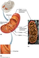
Cytoskeleton
The cytoskeleton is a network of filaments and tubules, including microtubules, microfilaments, and intermediate filaments. - Functions: Maintains cell shape, anchors organelles, assists in movement, and is dynamic. 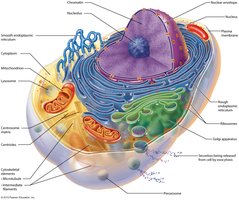
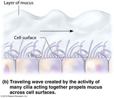
Cilia and Flagella
Extensions of the cell surface made of microtubules. - Cilia: Short, numerous; move fluids over cell surface (e.g., respiratory tract). - Flagella: Longer, fewer; move the cell itself (e.g., sperm cell). 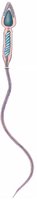
Microvilli
Microvilli are finger-shaped projections formed by plasma membrane folding, increasing surface area for absorption. 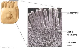
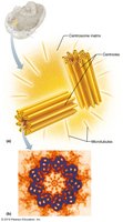
Centrioles and Centrosome
Centrosomes consist of two centrioles at right angles, essential for chromosome movement during cell division. 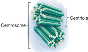
Plasma Membrane Structure and Function
Fluid Mosaic Model
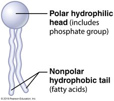
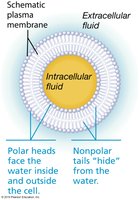
The plasma membrane is described by the Fluid Mosaic model, composed of phospholipids, proteins, cholesterol, glycolipids, and glycoproteins. - Phospholipid bilayer: Hydrophilic heads face water; hydrophobic tails face inward. 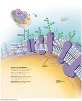
Additional Components
- Cholesterol: Maintains membrane fluidity. - Glycolipids and glycoproteins: Cell identification. - Proteins: Peripheral (surface) and integral (transmembrane) proteins with various functions.
Permeability of the Plasma Membrane
The plasma membrane is selectively permeable, allowing some substances to cross while restricting others. - Factors affecting permeability: Electric charge, molecule size, and lipid solubility.
Membrane Transport
Overview
Substances cross the plasma membrane via passive or active transport. - Passive transport: No energy required (diffusion, facilitated diffusion, osmosis). - Active transport: Energy required (primary and secondary active transport, vesicular transport).
Passive Transport: Diffusion
Diffusion is the movement of solute molecules from higher to lower concentration, driven by kinetic energy. 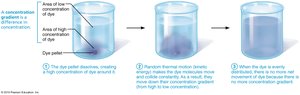
Simple Diffusion
Lipid-soluble molecules diffuse directly across the phospholipid bilayer. 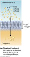
Facilitated Diffusion
Hydrophilic and large molecules require transmembrane proteins to cross the membrane. - Ion channels: Create tunnels for small ions. 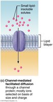 - Carrier proteins: Bind and transport larger molecules like glucose and amino acids.
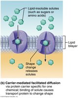
Osmosis
Osmosis is the diffusion of water across a selectively permeable membrane, driven by solute concentration. - Osmotic pressure: Force pulling water into a solution with higher solute concentration. 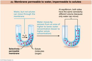
Tonicity
Tonicity describes the effect of extracellular solution on cell volume, based on non-penetrating solute concentration. - Hypertonic: Higher solute concentration outside; cells shrink. 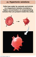 - Hypotonic: Lower solute concentration outside; cells swell.
 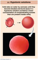 - Isotonic: Equal solute concentration; no change in cell volume.
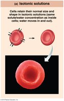
Osmolarity
Osmolarity measures the total solute concentration (penetrating and non-penetrating) in particles per liter. - Body fluid osmolarity: 300 mOsm. - Hyperosmotic: Higher osmolarity; hypertonic. - Hypoosmotic: Lower osmolarity; hypotonic.
Active Transport
Active transport requires energy (ATP) to move substances against their concentration gradient. - Pumps: Carrier proteins that transport ions and molecules.
Primary Active Transport
The Na+/K+ pump directly hydrolyzes ATP to move Na+ and K+ in opposite directions. 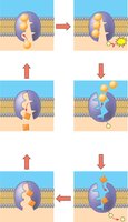
Secondary Active Transport
Uses energy stored in the Na+ gradient (created by the Na+/K+ pump) to transport other molecules, such as glucose, via SGLT (sodium-glucose cotransporter). 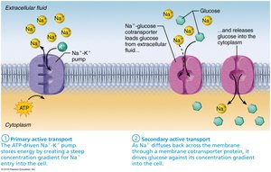
Vesicular Transport
Vesicular transport moves large molecules or groups of molecules in membrane sacs (vesicles), requiring ATP. - Endocytosis: Vesicles enter cells. - Exocytosis: Vesicles exit cells.
Phagocytosis
"Cell eating"; cell engulfs large particles. 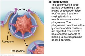
Pinocytosis
"Cell drinking"; cell engulfs fluid. 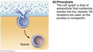
Summary Table: Cell Organelles and Functions
Organelle | Membrane? | Main Function |
|---|---|---|
Nucleus | Double | Stores DNA, controls cell |
Mitochondria | Double | ATP synthesis |
Ribosomes | No | Protein synthesis |
ER (Rough/Smooth) | Single | Protein/lipid synthesis |
Golgi apparatus | Single | Protein/lipid sorting |
Lysosomes | Single | Digestion |
Cytoskeleton | No | Structure, movement |
Cilia/Flagella | No | Movement |
Centrioles | No | Cell division |
Additional info: Academic context and explanations have been expanded for clarity and completeness.
