 Back
BackCells: The Living Units – Structure and Function
Study Guide - Smart Notes
 Tailored notes based on your materials, expanded with key definitions, examples, and context.
Cells: The Living Units
Introduction to Cells
Cells are the fundamental building blocks of all living organisms. According to the Cell Theory, all living things are composed of cells, and all cells arise from pre-existing cells. The human body contains approximately 50-100 trillion cells, but only about 10% are human; the rest are microbial, highlighting the complexity of our biological makeup.
General Structure of a Cell
Plasma Membrane: The outer boundary that separates the cell from its environment.
Cytoplasm: The internal fluid containing organelles and cytosol.
Nucleus: The control center housing genetic material.
Plasma Membrane: The Fluid Mosaic Model
Structure and Composition
The plasma membrane is a dynamic structure composed of a phospholipid bilayer with embedded proteins, cholesterol, and carbohydrates. It functions as a selective barrier, maintaining the internal environment of the cell.
Phospholipids: Form the basic structure, with hydrophilic heads facing outward and hydrophobic tails inward.
Cholesterol: Stabilizes membrane fluidity.
Glycolipids and Glycoproteins: Involved in cell recognition and signaling.
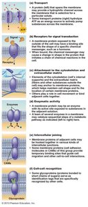
Proteins: Integral and peripheral proteins serve as channels, receptors, enzymes, and anchors.
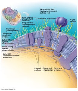
Functions of the Plasma Membrane
Mechanical Barrier: Separates intracellular and extracellular environments.
Selective Permeability: Regulates entry and exit of substances.
Electrochemical Gradient: Maintains ion gradients essential for cell function.
Communication: Contains receptors for signaling molecules.
Cell Signaling: Facilitates cellular responses to external stimuli.
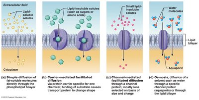
Movement of Substances Across the Membrane
Passive Transport
Passive transport does not require energy and relies on concentration gradients.
Diffusion: Movement of molecules from high to low concentration.
Facilitated Diffusion: Uses carrier or channel proteins for transport of larger or charged molecules.
Osmosis: Diffusion of water across a selectively permeable membrane.
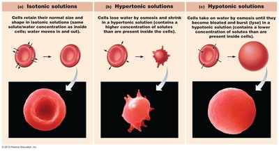
Tonicity
Tonicity describes the effect of extracellular solutions on cell volume.
Isotonic: No net water movement; cell retains normal shape.
Hypertonic: Water leaves the cell; cell shrinks (crenates).
Hypotonic: Water enters the cell; cell swells and may burst (lyse).
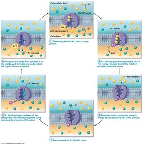
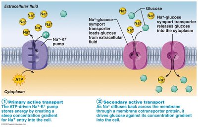
Active Transport
Active transport requires energy (usually ATP) to move substances against their concentration gradients.
Primary Active Transport: Direct use of ATP, e.g., sodium-potassium pump.
Secondary Active Transport: Indirect use of ATP; uses gradients established by primary transport.
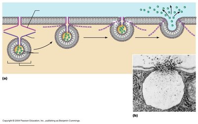
Vesicular Transport
Large particles and macromolecules are transported via vesicles, requiring ATP hydrolysis.
Exocytosis: Secretion of substances out of the cell.
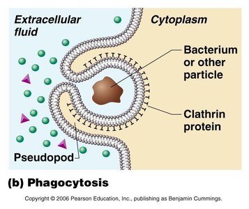
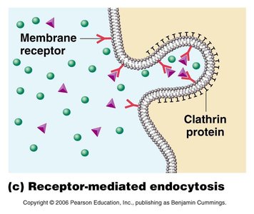
Endocytosis: Uptake of substances into the cell (includes phagocytosis, pinocytosis, and receptor-mediated endocytosis).
Transcytosis: Movement into, across, and out of the cell.
Vesicular Trafficking: Movement of substances within the cell.
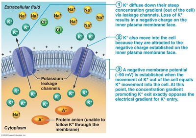
Membrane Potentials
Resting Membrane Potential
The resting membrane potential is the voltage difference across the plasma membrane due to unequal distribution of ions, primarily potassium (K+) and sodium (Na+).
Leaky Channels: Allow passive movement of ions, contributing to the potential.
Polarization: The inside of the cell is negatively charged relative to the outside.
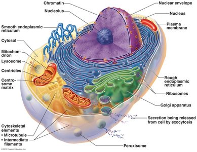
Cellular Organelles and Their Functions
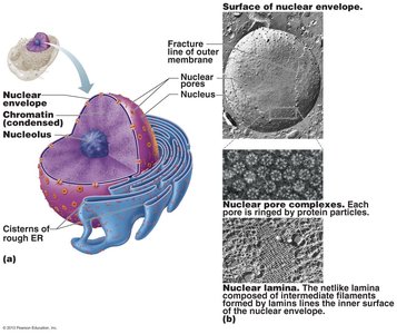
Nucleus
The nucleus is the control center of the cell, containing genetic material (DNA) and the nucleolus, where ribosomal RNA is synthesized.
Nuclear Envelope: Double membrane with nuclear pores for transport.
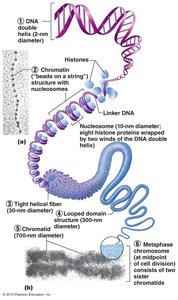
Chromatin: DNA wrapped around histones; condenses to form chromosomes during cell division.
Cytoplasm and Organelles
Cytosol: Fluid portion containing dissolved substances.
Inclusions: Stored nutrients, secretory products, and pigment granules.
Organelles: Specialized structures with distinct functions.
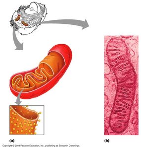
Mitochondria
Double-membraned organelles responsible for ATP production via aerobic respiration. The inner membrane forms cristae to increase surface area for energy production.
Ribosomes
Granules composed of protein and rRNA; sites of protein synthesis. Free ribosomes synthesize cytosolic proteins, while membrane-bound ribosomes produce proteins for membranes or export.
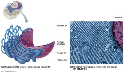
Endoplasmic Reticulum (ER)
Rough ER (rER): Studded with ribosomes; synthesizes proteins and phospholipids.
Smooth ER (sER): Lacks ribosomes; involved in lipid metabolism, detoxification, and calcium storage.
Golgi Apparatus
Stacked, flattened membranes that modify, sort, and package proteins and lipids for secretion or delivery to other organelles.
Vesicles, Lysosomes, and Peroxisomes
Vesicles: Membrane-bound sacs for transport within cells.
Lysosomes: Contain digestive enzymes for breaking down waste, pathogens, and cellular debris.
Peroxisomes: Contain oxidases and catalases for detoxifying harmful substances and neutralizing free radicals.
Cytoskeleton
A network of protein filaments (microtubules, microfilaments, intermediate filaments) that provide structural support, shape, and facilitate movement within the cell.
Protein Synthesis
From DNA to Protein
Protein synthesis involves two main processes: transcription and translation.
Transcription: In the nucleus, DNA is used as a template to synthesize messenger RNA (mRNA).
Translation: At the ribosome, mRNA is decoded to assemble a polypeptide chain using transfer RNA (tRNA) and ribosomal RNA (rRNA).
Transcription Steps
Initiation: RNA polymerase binds to the promoter region of DNA.
Elongation: RNA polymerase synthesizes the mRNA strand.
Termination: Synthesis ends at a termination signal; mRNA is released.
Translation Steps
Initiation: mRNA binds to the ribosome; tRNA brings the first amino acid.
Elongation: Ribosome moves along mRNA, adding amino acids to the growing chain.
Termination: Stop codon is reached; polypeptide is released.
Role of Rough ER in Protein Synthesis
The mRNA–ribosome complex is directed to the rough ER, where the growing protein enters the ER lumen, may be modified, and is then packaged into vesicles for transport to the Golgi apparatus for further processing.
Summary Table: Types of Membrane Transport
Process | Energy Source | Example |
|---|---|---|
Primary active transport | ATP | Pumping of ions across membranes |
Secondary active transport | Ion gradient | Movement of polar or charged solutes |
Exocytosis | ATP | Secretion of hormones/neurotransmitters |
Phagocytosis | ATP | White blood cell phagocytosis |
Pinocytosis | ATP | Absorption by intestinal cells |
Receptor-mediated endocytosis | ATP | Hormone and cholesterol uptake |
Additional info: This guide covers the essential structure and function of cells, focusing on the plasma membrane, organelles, and the processes of membrane transport and protein synthesis, as outlined in a typical Anatomy & Physiology college curriculum.
