 Back
BackCells: The Living Units – Structure, Function, and Processes
Study Guide - Smart Notes
 Tailored notes based on your materials, expanded with key definitions, examples, and context.
Cells: The Living Units.
Cell Theory and Diversity
The cell is the fundamental unit of life, forming the basis for all living organisms. Human bodies contain trillions of cells, with over 250 distinct types, each specialized for unique functions.
Cell Theory: All living things are composed of cells; cells are the smallest units of life; all cells arise from pre-existing cells.
Cell Diversity: Cells vary in size, shape, and function, reflecting their specialized roles in the body.
Extracellular Materials
Substances found outside cells are essential for cellular function and communication.
Extracellular Fluids (ECFs): Include interstitial fluid, blood plasma, and cerebrospinal fluid.
Cellular Secretions: Such as saliva, mucus, and gastric fluids, facilitate various physiological processes.
Extracellular Matrix: A network of proteins and polysaccharides that provides structural support and mediates cell signaling.
Plasma Membrane Structure and Function
Fluid Mosaic Model
The plasma membrane is a dynamic, selectively permeable barrier composed of a phospholipid bilayer with embedded proteins and carbohydrates.
Phospholipids: Form the basic structure, with hydrophilic heads facing outward and hydrophobic tails inward.
Cholesterol: Stabilizes membrane fluidity and integrity.
Proteins: Integral and peripheral proteins serve as channels, receptors, enzymes, and anchors.
Glycocalyx: Carbohydrate-rich area on the cell surface, important for cell recognition and immune response.
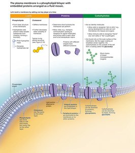
Membrane Lipids and Proteins
Phospholipid Bilayer: Provides the basic structure and barrier to water-soluble substances.
Integral Proteins: Span the membrane and are involved in transport and signaling.
Peripheral Proteins: Attach to membrane surfaces and assist in cell signaling and structure.

Glycocalyx
The glycocalyx is a carbohydrate-rich coating on the cell surface, functioning as a biological marker for cell recognition and immune interactions.
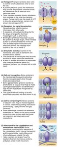
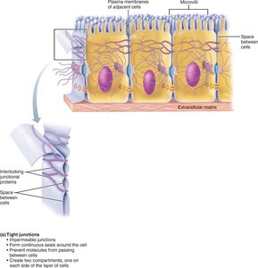
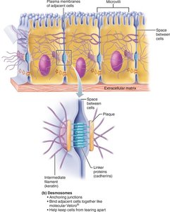
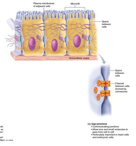
Intercellular Junctions
Cells are connected by specialized junctions that maintain tissue integrity and facilitate communication.
Tight Junctions: Prevent passage of substances between cells.
Desmosomes: Anchor cells together, providing mechanical stability.
Gap Junctions: Allow direct communication between cells via channels.
Membrane Transport Mechanisms
Passive Transport
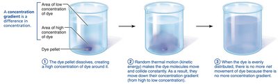
Passive transport moves substances across the membrane without energy input, driven by concentration gradients.
Simple Diffusion: Movement of lipid-soluble molecules directly through the bilayer.
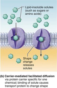
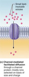
Facilitated Diffusion: Movement of molecules via protein carriers or channels (e.g., glucose, ions).
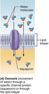
Osmosis: Diffusion of water through a selectively permeable membrane.
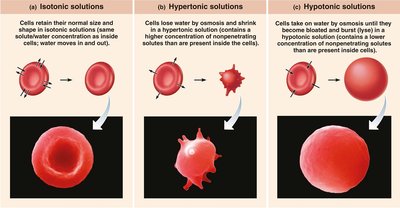
Osmolarity and Tonicity
Osmolarity refers to the total solute concentration in a solution, while tonicity describes a solution's effect on cell volume.
Isotonic: No net water movement; cell retains normal shape.
Hypertonic: Water leaves the cell; cell shrinks (crenates).
Hypotonic: Water enters the cell; cell swells and may lyse.
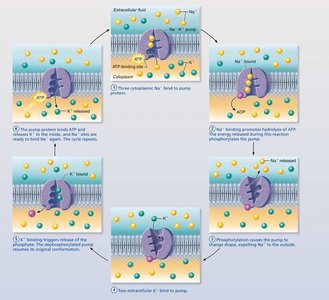
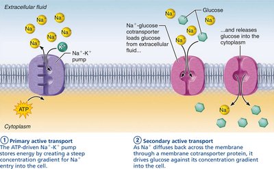
Active Transport
Active transport requires energy (ATP) to move substances against their concentration gradients.
Primary Active Transport: Direct use of ATP (e.g., Na+-K+ pump).
Secondary Active Transport: Uses energy stored in ion gradients created by primary active transport.
Vesicular Transport
Vesicular transport moves large particles and fluids across membranes via vesicles, requiring ATP.
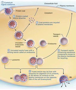
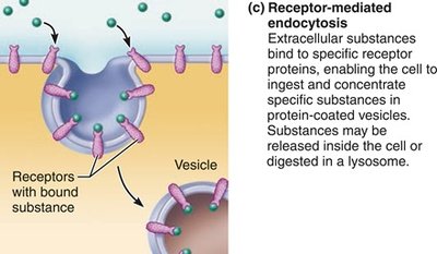
Endocytosis: Uptake of materials (phagocytosis, pinocytosis, receptor-mediated endocytosis).
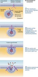
Exocytosis: Expulsion of materials from the cell.
Transcytosis: Transport into, across, and out of the cell.
Vesicular Trafficking: Movement of substances within the cell.
Membrane Potentials and Cell Communication
Resting Membrane Potential (RMP)
All cells maintain a voltage across their plasma membrane, typically -50 to -70 mV, due to the differential distribution of ions, especially K+ and Na+.
Cell Adhesion Molecules (CAMs) and Receptors
CAMs anchor cells, assist in movement, and mediate immune responses. Plasma membrane receptors are involved in chemical and contact signaling, often using G proteins as second messengers.
Cytoplasm and Organelles
Cytoplasm
The cytoplasm is the cellular material between the plasma membrane and nucleus, containing cytosol, inclusions, and organelles.
Cytosol: Fluid portion with dissolved substances.
Inclusions: Stored nutrients and pigments.
Organelles: Specialized structures performing cellular functions.
Major Organelles
Mitochondria: Site of ATP production via aerobic respiration; contain their own DNA and replicate by fission.
Ribosomes: Sites of protein synthesis; free in cytosol or bound to rough ER.
Endoplasmic Reticulum (ER): Rough ER synthesizes proteins; smooth ER synthesizes lipids, detoxifies chemicals, and stores calcium.
Golgi Apparatus: Modifies, sorts, and packages proteins and lipids for secretion or delivery to other organelles.
Lysosomes: Contain digestive enzymes for breakdown of waste and cellular debris.
Peroxisomes: Detoxify harmful substances and metabolize fatty acids.
Cytoskeleton: Network of protein filaments providing structural support, cell shape, and movement.
Centrosome and Centrioles: Organize microtubules and are essential for cell division.
Cilia and Microvilli: Cilia move substances across cell surfaces; microvilli increase surface area for absorption.
Nucleus and Genetic Material
Nucleus Structure
The nucleus is the control center of the cell, containing genetic material and directing cellular activities.
Nuclear Envelope: Double membrane with pores for molecular exchange.
Nucleoli: Sites of ribosomal RNA synthesis and ribosome assembly.
Chromatin: DNA-protein complex; condenses to form chromosomes during cell division.
Cell Cycle and Division
Phases of the Cell Cycle
The cell cycle consists of interphase (G1, S, G2) and the mitotic phase (mitosis and cytokinesis).
Interphase: Cell growth, DNA replication, and preparation for division.
Mitosis: Division of the nucleus (prophase, metaphase, anaphase, telophase).
Cytokinesis: Division of the cytoplasm, resulting in two daughter cells.
Control of Cell Division
Cell division is regulated by internal and external signals, including growth factors, cell size, and contact inhibition. Checkpoints ensure proper division; failure can lead to uncontrolled growth (cancer).
Cell Death and Renewal
Autophagy, Proteasomes, and Apoptosis
Autophagy: Removal of damaged organelles via lysosomal degradation.
Proteasomes: Degrade misfolded or unneeded proteins tagged with ubiquitin.
Apoptosis: Programmed cell death, essential for development and homeostasis.
Stem and Progenitor Cells
Types and Functions
Stem Cells: Undifferentiated cells capable of self-renewal and differentiation into various cell types.
Totipotent: Can become any cell type (early embryo).
Pluripotent: Can become most cell types (embryonic stem cells).
Multipotent: Can become a limited range of cells (adult stem cells).
Progenitor Cells: More specialized than stem cells; can be unipotent (one cell type) or oligopotent (few cell types).
Comparison Table: Stem Cells vs. Progenitor Cells
Feature | Stem Cells | Progenitor Cells |
|---|---|---|
Potency | Totipotent, pluripotent, multipotent | Unipotent, oligopotent |
Self-renewal | Indefinite | Limited |
Differentiation | Many cell types | Few cell types |
Activation | Development, repair | Usually inactive, activated by damage |
Cell Differentiation, Growth, and Aging
Cell Differentiation
Cells develop specialized structures and functions through differentiation, guided by gene expression and environmental cues.
Cell Growth and Atrophy
Hyperplasia: Increased cell number due to accelerated growth.
Atrophy: Decreased cell size from loss of stimulation or use.
Cell Aging
Cellular aging is influenced by genetic, environmental, and metabolic factors. Theories include wear and tear, mitochondrial dysfunction, immune decline, and genetic programming (telomere shortening).
Telomeres: Protective DNA sequences at chromosome ends; shorten with each division.
Telomerase: Enzyme that extends telomeres, active in germ cells and cancer cells.
