Back
BackCellular Anatomy & Physiology: The Synthesis Story
Study Guide - Smart Notes
 Tailored notes based on your materials, expanded with key definitions, examples, and context.
The Living Units: The Synthesis Story
Introduction to Cell Anatomy & Cellular Pathways
This section provides a comprehensive overview of cell structure, function, and the pathways that drive cellular activity. Understanding these foundational concepts is essential for mastering anatomy and physiology at the college level.
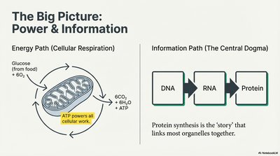
The Big Picture: Power & Information
Energy Path (Cellular Respiration) & Information Path (Central Dogma)
Cellular Respiration: Cells convert glucose and oxygen into carbon dioxide, water, and ATP (energy currency of the cell).
Central Dogma: Information flows from DNA to RNA to Protein, guiding cellular structure and function.
Protein Synthesis: Links organelles and is central to cell function.
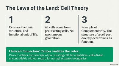
The Laws of the Land: Cell Theory
Three Fundamental Principles
Cells are the basic structural and functional unit of life.
All cells come from pre-existing cells. No spontaneous generation occurs.
Principle of Complementarity: Structure determines function.
Clinical Connection: Cancer violates cell theory by dividing uncontrollably, ignoring normal regulatory boundaries.
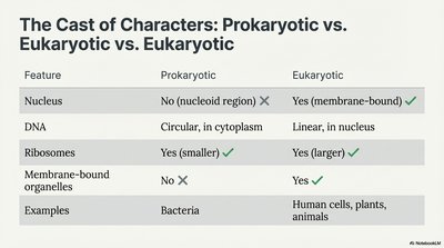
The Cast of Characters: Prokaryotic vs. Eukaryotic Cells
Key Differences
Feature | Prokaryotic | Eukaryotic |
|---|---|---|
Nucleus | No (nucleoid region) | Yes (membrane-bound) |
DNA | Circular, in cytoplasm | Linear, in nucleus |
Ribosomes | Yes (smaller) | Yes (larger) |
Membrane-bound organelles | No | Yes |
Examples | Bacteria | Human cells, plants, animals |
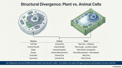
Structural Divergence: Plant vs. Animal Cells
Major Structural Differences
Feature | Animal | Plant |
|---|---|---|
Cell Wall | No | Yes (cellulose) |
Central Vacuole | No | Yes (large, provides turgor) |
Shape | Round | Rectangular |
Energy Organelles | Mitochondria | Mitochondria & chloroplasts |
Centrioles | Yes | No |
Lysosomes | Yes | Rare |
Key Takeaway: Structural differences explain why plants can stand tall and animals can move and flex.
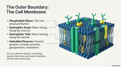
The Outer Boundary: The Cell Membrane
Structure and Function
Phospholipid Bilayer: Main structural barrier.
Hydrophilic Heads: Face outward, water-loving.
Hydrophobic Tails: Face inward, water-fearing.
Embedded Elements: Channel proteins, receptor proteins, glycoproteins, cholesterol.
Acts as a selective barrier, controlling entry and exit of substances to maintain homeostasis.
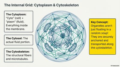
The Internal Grid: Cytoplasm & Cytoskeleton
Organization and Support
Cytoplasm: Everything inside the membrane (cytosol + organelles).
Cytosol: The fluid portion.
Cytoskeleton: Network of fibers and microtubules for support and transport.
Key Concept: Organelles are anchored and transported along the cytoskeleton, not floating randomly.
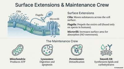
Surface Extensions & Maintenance Crew
Specialized Structures and Organelles
Cilia: Move substances across the cell surface.
Flagella: Propel the entire cell (e.g., sperm).
Microvilli: Increase surface area for absorption.
Mitochondria: Produce ATP.
Lysosomes: Digest and recycle cellular waste.
Peroxisomes: Detoxify harmful substances.
Smooth ER: Synthesizes lipids and carbohydrates.
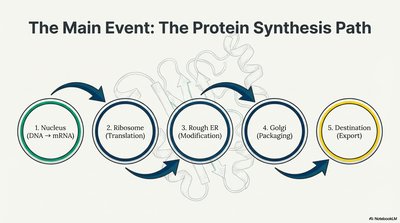
The Main Event: The Protein Synthesis Path
Steps in Protein Synthesis
Nucleus: DNA is transcribed to mRNA.
Ribosome: mRNA is translated into a polypeptide chain.
Rough ER: Protein is modified.
Golgi Apparatus: Protein is packaged.
Destination: Protein is exported or used within the cell.
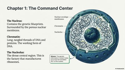
Chapter 1: The Command Center (Nucleus)
Structure and Function
Nucleus: Contains genetic material, surrounded by a nuclear envelope.
Chromatin: DNA-protein complex, the working form of DNA.
Nucleolus: Site of ribosome production.
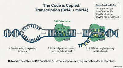
The Code is Copied: Transcription (DNA → mRNA)
Mechanism of Transcription
DNA unwinds, exposing bases.
RNA polymerase reads the template strand.
mRNA is synthesized as a complementary strand.
Outcome: Mature mRNA exits the nucleus, carrying instructions for one protein.
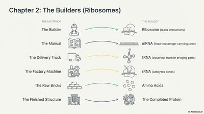
Chapter 2: The Builders (Ribosomes)
Roles in Protein Synthesis
Ribosome: Reads mRNA instructions.
mRNA: Messenger carrying the code.
tRNA: Transfers amino acids to the ribosome.
rRNA: Catalyzes peptide bond formation.
Amino Acids: Raw materials for proteins.
Completed Protein: Final product of translation.
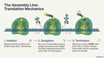
The Assembly Line: Translation Mechanics
Steps of Translation
Initiation: Ribosome binds to mRNA at the start codon (AUG).
Elongation: tRNA brings amino acids, chain grows.
Termination: Ribosome reaches a stop codon, protein is released.
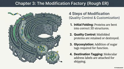
Chapter 3: The Modification Factory (Rough ER)
Protein Quality Control & Customization
Initial Folding: Proteins bent into correct 3D shapes.
Quality Control: Misfolded proteins are retained or destroyed.
Glycosylation: Addition of sugar tags for function.
Destination Tagging: Address labels for shipping.
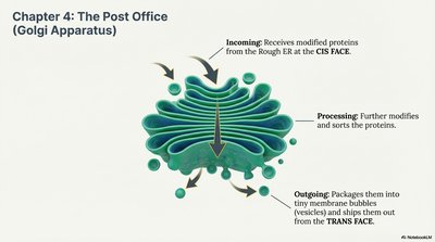
Chapter 4: The Post Office (Golgi Apparatus)
Processing and Shipping Proteins
Incoming: Receives proteins from Rough ER (cis face).
Processing: Further modifies and sorts proteins.
Outgoing: Packages proteins into vesicles for export (trans face).
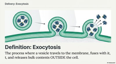
Delivery: Exocytosis
Definition and Mechanism
Exocytosis: The process where a vesicle travels to the membrane, fuses with it, and releases bulk contents outside the cell.
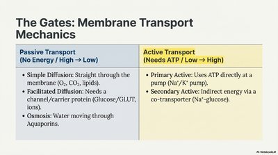
The Gates: Membrane Transport Mechanics
Passive vs. Active Transport
Passive Transport (No Energy / High → Low) | Active Transport (Needs ATP / Low → High) | |
|---|---|---|
Simple Diffusion | Straight through membrane (O2, CO2, lipids) | |
Facilitated Diffusion | Needs channel/carrier protein (glucose, ions) | |
Osmosis | Water through aquaporins | |
Primary Active | Uses ATP directly (Na+/K+ pump) | |
Secondary Active | Indirect energy via co-transporter (Na+-glucose) |
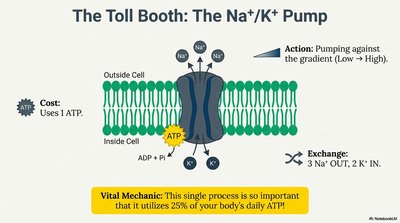
The Toll Booth: The Na+/K+ Pump
Mechanism and Importance
Action: Pumps 3 Na+ out, 2 K+ in, against the gradient.
Cost: Uses 1 ATP per cycle.
Vital Mechanic: Utilizes 25% of the body's daily ATP.
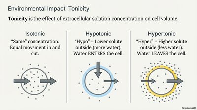
Environmental Impact: Tonicity
Effect of Extracellular Solution on Cell Volume
Isotonic: Equal solute concentration; no net water movement.
Hypotonic: Lower solute outside; water enters cell.
Hypertonic: Higher solute outside; water leaves cell.
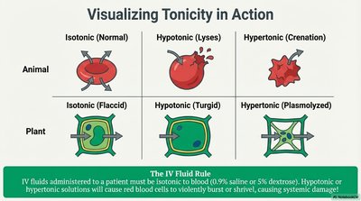
Visualizing Tonicity in Action
Effects on Animal and Plant Cells
Isotonic | Hypotonic | Hypertonic | |
|---|---|---|---|
Animal | Normal | Lyses (bursts) | Crenation (shrivels) |
Plant | Flaccid | Turgid | Plasmolyzed |
The IV Fluid Rule: IV fluids must be isotonic to blood to prevent cell damage.