 Back
BackCellular Level of Organization: Structure and Function in Human Anatomy & Physiology
Study Guide - Smart Notes
 Tailored notes based on your materials, expanded with key definitions, examples, and context.
Chapter 3: The Cellular Level of Organization
Introduction to Cells
Cells are the fundamental units of life in the human body, forming the basis for all physiological functions. Understanding cell structure and function is essential for comprehending higher levels of biological organization.
Definition: Cells are the smallest living units in the human body, classified as eukaryotic cells.
Building Blocks: All organisms are composed of cells, which arise from the division of preexisting cells.
Homeostasis: Each cell maintains homeostasis at the cellular level, contributing to tissue, organ, and organismal homeostasis.
Cell Numbers: The human body contains trillions of cells, with even more microbial cells present.
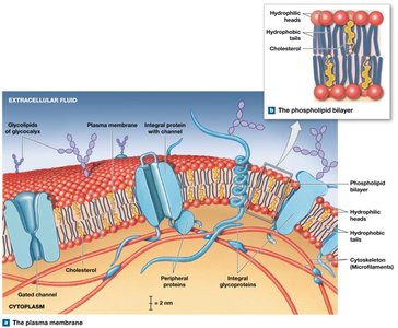
Plasma Membrane Structure and Function
The plasma membrane forms the boundary of the cell, regulating the passage of substances and maintaining distinct internal and external environments.
Composition: Major components include phospholipids and cholesterol.
Functions: Physical separation, regulation of transport, sensitivity (cell communication), and support (anchoring cells).
Phospholipid Bilayer: Consists of hydrophilic heads (water-loving) facing the extracellular and intracellular fluids, and hydrophobic tails (water-fearing) forming the interior.
Cholesterol: Stiffens the membrane, reducing fluidity and permeability.
Proteins: Integral (embedded), transmembrane (span the membrane), and peripheral (surface-bound) proteins perform various functions.
Eukaryotic Cell Organelles
Organelles are specialized structures within cells that perform distinct functions, contributing to cellular homeostasis and activity.
Cytoplasm: All material between the plasma membrane and nuclear envelope, including cytosol, organelles, and inclusions.
Cytosol: Jelly-like fluid containing water, nutrients, ions, proteins, and waste.
Inclusions: Insoluble masses such as glycogen granules, lipid droplets, and pigment granules.
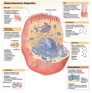
Nonmembranous Organelles
Cytoskeleton: Internal protein framework providing strength and flexibility. Types include microfilaments (actin), intermediate filaments, and microtubules (tubulin).
Centrioles: Cylindrical structures involved in spindle formation during cell division.
Ribosomes: Sites of protein synthesis, composed of rRNA and proteins. Free ribosomes synthesize proteins for cytosol; fixed ribosomes (on RER) synthesize proteins for export or organelles.
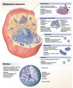
Membranous Organelles
Endoplasmic Reticulum (ER): Network of membranes continuous with the nuclear envelope. Smooth ER (SER) synthesizes lipids, steroids, and stores ions; Rough ER (RER) has ribosomes and synthesizes proteins.
Golgi Apparatus: Modifies, packages, and sorts proteins and lipids for secretion or use within the cell.
Lysosomes: Vesicles containing digestive enzymes for hydrolyzing polymers, recycling organelles, and autolysis.
Peroxisomes: Vesicles containing enzymes for breaking down fatty acids and detoxifying chemicals.
Mitochondria: Sites of ATP production; number varies by cell type according to energy demand.
Nucleus: Control center storing genetic information (DNA), directing protein synthesis, and determining cell structure and function.
Cytoskeleton
The cytoskeleton is a dynamic network of protein fibers that provides structural support, facilitates movement, and organizes cellular components.
Microfilaments: Smallest fibers, composed of actin; provide mechanical strength and participate in muscle contraction.
Intermediate Filaments: Provide structural stability and maintain cell shape.
Microtubules: Largest fibers, hollow tubes of tubulin; radiate from the centrosome, anchor organelles, and form spindle apparatus during cell division.

Protein Synthesis: Transcription and Translation
Protein synthesis is the process by which cells produce proteins, following the instructions encoded in DNA. This involves two main steps: transcription and translation.
Transcription: DNA nucleotide sequence is copied to mRNA in the nucleus. Introns are removed, and exons are spliced together during RNA processing.
Translation: mRNA binds to ribosomes in the cytoplasm. tRNA delivers amino acids, matching anticodons to mRNA codons, forming a polypeptide chain.
Central Dogma:
Genetic Code: Each codon (three bases) specifies an amino acid.
Transport Across the Plasma Membrane
Cells regulate the movement of substances across the plasma membrane using various transport mechanisms, ensuring proper internal conditions.
Selective Permeability: The plasma membrane allows some substances to pass freely while restricting others, based on size, charge, shape, and solubility.
Passive Transport: No energy required; includes diffusion and osmosis.
Active Transport: Requires energy (ATP); moves substances against concentration gradients.
Diffusion
Definition: Net movement from higher to lower concentration.
Simple Diffusion: Lipid-soluble compounds and gases pass directly through the membrane.
Channel-Mediated Diffusion: Small water-soluble compounds and ions pass through protein channels.
Osmosis
Definition: Net diffusion of water across a selectively permeable membrane in response to solute concentration differences.
Direction: Water moves toward higher solute concentration (lower water concentration).
Equilibrium: Osmosis continues until solute concentrations are equal on both sides.
Tonicity
Isotonic: Equal solute concentrations; cell size remains unchanged.
Hypotonic: Lower solute concentration outside; water enters cell, causing swelling and possible bursting (hemolysis).
Hypertonic: Higher solute concentration outside; water exits cell, causing shrinkage (crenation).
Carrier-Mediated Transport
Carrier Proteins: Specialized integral proteins that transport specific substances.
Symporters: Move two substances in the same direction.
Antiporters: Move two substances in opposite directions.
Facilitated Diffusion: Passive transport of large, water-soluble molecules via carrier proteins.
Active Transport: Uses ATP to move substances against gradients; includes ion pumps and exchange pumps (e.g., sodium-potassium ATPase).
Sodium-Potassium Pump Equation: (3 Na+ out, 2 K+ in per ATP)
Cell Life Cycle
Cell division is essential for growth, repair, and maintenance. The cell cycle includes interphase, mitosis, and cytokinesis.
Interphase: Period between divisions; includes G1 (growth), S (DNA replication), and G2 (protein synthesis).
G0 Phase: Nondividing state; some cells (e.g., neurons) remain here indefinitely.
Mitosis: Nuclear division; stages include prophase, metaphase, anaphase, and telophase.
Cytokinesis: Division of cytoplasm; cleavage furrow forms, separating daughter cells.
DNA Replication: Occurs in S phase; ensures each daughter cell receives a complete genome.
Mitosis Stages
Prophase: Chromatin condenses, nuclear envelope disintegrates, spindle fibers form.
Metaphase: Chromatid pairs align at metaphase plate.
Anaphase: Chromatids separate and move to opposite poles.
Telophase: Nuclear membranes reform, chromosomes return to chromatin, spindle breaks down.
Cytokinesis: Contractile band of microfilaments forms cleavage furrow, separating cells.
Example: Skin cells undergo frequent mitosis to replace damaged or lost cells, while neurons rarely divide after maturity.
