 Back
BackCentral Nervous System Structure and Function Study Guide
Study Guide - Smart Notes
 Tailored notes based on your materials, expanded with key definitions, examples, and context.
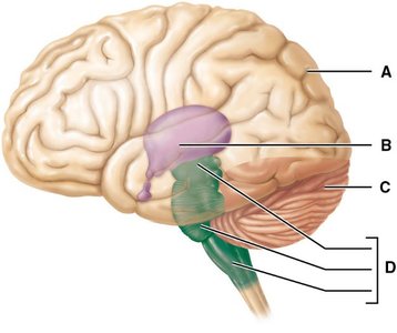
Q1. Name the four adult brain regions labeled A–D.
Background
Topic: Major Regions of the Adult Brain
This question tests your ability to identify the main anatomical regions of the adult brain using a labeled diagram. Understanding these regions is foundational for studying brain structure and function in anatomy and physiology (ANP).
Key Terms:
Cerebrum: The largest part of the brain, responsible for higher brain functions such as thought and action.
Diencephalon: Contains structures such as the thalamus and hypothalamus, involved in sensory and autonomic functions.
Brain Stem: Connects the brain to the spinal cord and controls vital life functions.
Cerebellum: Coordinates voluntary movements and balance.
Step-by-Step Guidance
Observe the diagram and note the location of each labeled region (A–D). Each label points to a distinct anatomical area of the brain.
Recall the general positions of the four major brain regions:
The cerebrum is the largest, most superior part.
The diencephalon is located centrally, deep to the cerebrum.
The brain stem is inferior to the diencephalon and anterior to the cerebellum.
The cerebellum is posterior and inferior, beneath the cerebrum and behind the brain stem.
Match each label (A–D) to the correct brain region based on its position in the diagram.
Write the name of each region next to the corresponding letter.
Try solving on your own before revealing the answer!
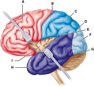
Q2. Identify the lobes and anatomical landmarks of the cerebral hemispheres labeled A–I.
Background
Topic: Lobes and Landmarks of the Cerebral Cortex
This question assesses your ability to recognize and name the major lobes and anatomical features of the cerebral hemispheres using a labeled diagram. This is essential for understanding brain localization of function.
Key Terms:
Lobes: Frontal, parietal, temporal, occipital (main divisions of the cerebrum).
Landmarks: Central sulcus, lateral sulcus, precentral gyrus, postcentral gyrus, etc.
Step-by-Step Guidance
Examine the diagram and locate each label (A–I). Each points to a specific lobe or anatomical landmark.
Recall the general locations of the cerebral lobes:
Frontal lobe: anterior portion
Parietal lobe: superior and posterior to the frontal lobe
Temporal lobe: lateral sides
Occipital lobe: posterior portion
Identify major sulci and gyri, such as the central sulcus (separates frontal and parietal lobes) and the lateral sulcus (separates temporal from frontal/parietal lobes).
Assign the correct lobe or landmark name to each label based on its position.
Try solving on your own before revealing the answer!
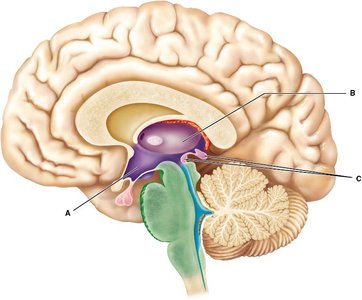
Q3. Name the structures of the diencephalon labeled A–C.
Background
Topic: Diencephalon Anatomy
This question focuses on identifying the three main subdivisions of the diencephalon, which are crucial for sensory relay, autonomic control, and endocrine function.
Key Terms:
Thalamus: Relay station for sensory information.
Hypothalamus: Regulates autonomic and endocrine functions.
Epithalamus: Includes the pineal gland, involved in circadian rhythms.
Step-by-Step Guidance
Look at the diagram and note the location of each label (A–C) within the central part of the brain.
Recall the relative positions:
Thalamus: centrally located, egg-shaped structure
Hypothalamus: inferior to the thalamus, near the base of the brain
Epithalamus: dorsal/posterior to the thalamus, often associated with the pineal gland
Assign the correct name to each labeled structure based on its position and shape.
Try solving on your own before revealing the answer!
