 Back
BackCh. 12 Central Nervous System: Structure, Development, and Major Brain Regions
Study Guide - Smart Notes
 Tailored notes based on your materials, expanded with key definitions, examples, and context.
Central Nervous System (CNS)
Overview and Functions
The central nervous system (CNS) is composed of the brain and spinal cord. It is responsible for integrating sensory information and accordingly. The CNS provides two-way communication between the brain and the body and serves as the major reflex center, where reflexes are initiated and completed at the spinal cord.
Cephalization: Evolutionary development of the anterior portion of the CNS, resulting in an increased number of neurons, with the highest level in the human brain.
Brain Development
Embryological Development
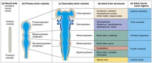
Embryologically, the brain and spinal cord begin as a neural tube. The anterior end of the neural tube expands and forms three primary brain vesicles, which further differentiate into five secondary brain vesicles and eventually into the adult brain structures.
Primary brain vesicles: Prosencephalon (forebrain), Mesencephalon (midbrain), Rhombencephalon (hindbrain)
Secondary brain vesicles: Telencephalon, Diencephalon, Mesencephalon, Metencephalon, Myelencephalon
Adult brain structures: Cerebrum, diencephalon (thalamus, hypothalamus, epithalamus), midbrain, pons, cerebellum, medulla oblongata, spinal cord
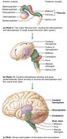
Folding and Growth of the Brain
The brain grows faster than the surrounding membranous skull, causing it to fold to occupy available space. The forebrain moves toward the brain stem, and the cerebral hemispheres envelop the diencephalon and midbrain, increasing surface area through creasing and folding.


Brain Regions and Organization
Major Regions of the Adult Brain
Cerebral Hemispheres: Includes the cerebral cortex (gray matter), white matter, and basal nuclei
Diencephalon: Thalamus, hypothalamus, epithalamus
Brain Stem: Midbrain, pons, medulla oblongata
Cerebellum
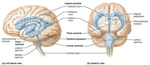
Ventricles of the Brain
Structure and Function
The ventricles are fluid-filled chambers within the brain, continuous with each other and the central canal of the spinal cord. They are filled with cerebrospinal fluid (CSF) and lined by ependymal cells. The paired lateral ventricles are separated by the septum pellucidum and connect to the third ventricle via the interventricular foramen. The third ventricle connects to the fourth ventricle via the cerebral aqueduct. The fourth ventricle is continuous with the central canal and connects to the subarachnoid space via three openings.
Cerebral Hemispheres
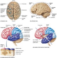
Surface Features and Lobes
The cerebral hemispheres form the superior part of the brain and account for 83% of its mass. They are connected by the corpus callosum. Surface markings include:
Gyri: Ridges
Sulci: Shallow grooves
Fissures: Deep grooves (e.g., longitudinal fissure, transverse cerebral fissure)
Each hemisphere is divided into five lobes: frontal, parietal, temporal, occipital, and insula (the latter is buried under portions of the other lobes).

Cerebral Cortex
Structure and Functional Areas
The cerebral cortex is the "executive suite" of the brain, responsible for conscious mind functions such as awareness, sensory perception, voluntary motor initiation, communication, memory storage, and understanding. It is a thin (2–4 mm) superficial layer of gray matter, making up 40% of the brain's mass.
Contains three types of functional areas: motor areas (voluntary movement), sensory areas (conscious awareness of sensation), and association areas (integration of information).
Each hemisphere controls the contralateral side of the body.
Lateralization of cortical function can occur in only one hemisphere.
Conscious behavior involves the entire cortex.
Diencephalon
Major Structures
The diencephalon consists of three paired gray matter structures: thalamus, hypothalamus, and epithalamus. All three enclose the third ventricle.
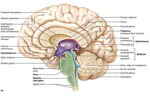
Thalamus: Major sensory relay station and gateway to the cerebral cortex; consists of bilateral egg-shaped nuclei connected by the interthalamic adhesion.
Hypothalamus: Located below the thalamus; main visceral control and regulating center vital to homeostasis. Contains mammillary bodies (olfactory relay stations) and the infundibulum (connects to pituitary gland).
Epithalamus: Most dorsal portion; contains the pineal gland, which secretes melatonin to regulate the sleep-wake cycle.
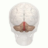
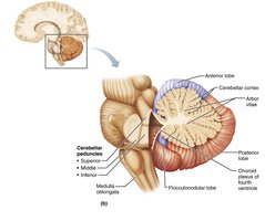
Cerebellum
Structure and Function
The cerebellum accounts for about 11% of brain mass and is located dorsal to the pons and medulla. It processes input from the cortex, brain stem, and sensory receptors to provide precise, coordinated movements of skeletal muscles and plays a major role in balance.
Cerebellar hemispheres are connected by the vermis.
Folia are transversely oriented gyri.
Each hemisphere has three lobes: anterior, posterior, and flocculonodular.
Contains a thin cortex of gray matter with a distinctive treelike pattern of white matter called the arbor vitae.
Purkinje cells originate in the cortex and synapse with the cerebellum.
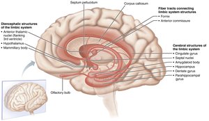
Limbic System
Structure and Function
The limbic system is a group of structures on the medial aspects of the cerebral hemispheres and diencephalon. It is involved in emotion, motivation, and memory formation.
Amygdaloid body: Recognizes angry or fearful facial expressions, assesses danger, and elicits fear responses.
Fornix: Fiber tract linking limbic system regions.
Cingulate gyrus: Plays a role in expressing emotions via gestures and resolving mental conflict.
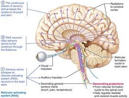
Reticular Formation
Structure and Function
The reticular formation extends through the central core of the brain stem and consists of three broad columns of nuclei. It has widespread axonal connections with the hypothalamus, thalamus, cerebral cortex, cerebellum, and spinal cord, allowing it to govern brain arousal.
Reticular activating system (RAS): Sends impulses to the cerebral cortex to keep it conscious and alert; filters out repetitive, familiar, or weak stimuli.
Inhibited by sleep centers, alcohol, and drugs; severe injury can result in coma.
Motor function helps control coarse limb movements via reticulospinal tracts.
Regulates visceral motor functions (vasomotor, cardiac, and respiratory centers).
Summary Tables: Functions of Major Brain Regions
Cerebral Hemispheres, Diencephalon, Brain Stem, and Cerebellum
The following tables summarize the main functions of the major brain regions. (Tables are referenced from the images and described below.)
Region | Main Functions |
|---|---|
Cerebral Hemispheres | Conscious thought, voluntary movement, interpretation of sensory input, memory, and learning |
Diencephalon | Relay and processing centers for sensory information, autonomic functions, hormone production |
Brain Stem | Conduction pathway between higher and lower brain centers; controls autonomic behaviors essential for survival |
Cerebellum | Coordinates voluntary movements, balance, and posture |
*Additional info: For more detailed breakdowns, refer to the labeled diagrams and tables in the source material, which provide specific nuclei and their functions within each region.*
Spinal Cord
Structure and Function
The spinal cord is enclosed in the vertebral column, beginning at the foramen magnum and ending at the level of the first or second lumbar vertebra. It provides two-way communication between the brain and body and serves as the major reflex center.
