 Back
BackCentral Nervous System: Structure, Function, and Protection
Study Guide - Smart Notes
 Tailored notes based on your materials, expanded with key definitions, examples, and context.
Central Nervous System: Embryonic Development
Overview of CNS Development
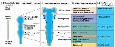
The central nervous system (CNS) develops from the neural tube during embryogenesis. This process establishes the foundational structures of the adult brain and spinal cord.
Neural Tube: The precursor to the CNS, containing the neural canal.
Primary Brain Vesicles: Prosencephalon (forebrain), Mesencephalon (midbrain), Rhombencephalon (hindbrain).
Secondary Brain Vesicles: Further differentiation leads to the formation of the telencephalon, diencephalon, mesencephalon, metencephalon, and myelencephalon.
Adult Brain Structures: These vesicles give rise to the cerebrum, diencephalon, brain stem (midbrain, pons, medulla oblongata), cerebellum, and spinal cord.
Neural Canal Regions: Develop into the lateral ventricles, third ventricle, cerebral aqueduct, fourth ventricle, and central canal.
Regions and Organization of the CNS
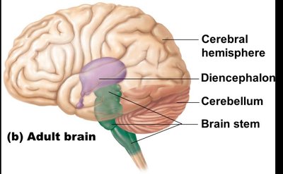
Major Brain Regions
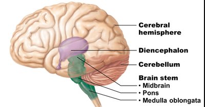
The adult brain is organized into four main regions, each with distinct functions and anatomical features.
Cerebral Hemispheres: The largest part of the brain, responsible for higher cognitive functions.
Diencephalon: Contains the thalamus, hypothalamus, and epithalamus; involved in sensory relay and homeostasis.
Brain Stem: Composed of the midbrain, pons, and medulla oblongata; controls vital autonomic functions.
Cerebellum: Coordinates movement and balance.
Gray and White Matter Organization
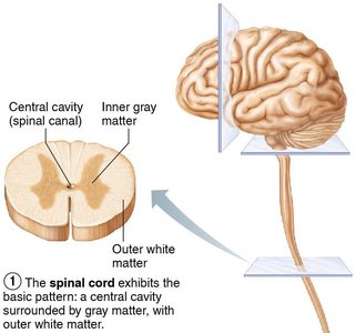
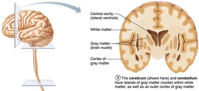
The CNS exhibits a characteristic pattern of gray and white matter distribution.
Spinal Cord: Central cavity surrounded by gray matter, with external white matter composed of myelinated fiber tracts.
Brain: Similar pattern, but with additional gray matter nuclei (basal nuclei) and an outer cortex of gray matter in the cerebrum and cerebellum.
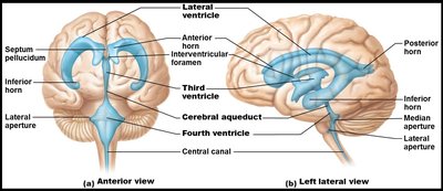
Ventricles of the Brain
Structure and Function
The brain contains interconnected cavities called ventricles, filled with cerebrospinal fluid (CSF) and lined by ependymal cells.
Lateral Ventricles: Paired, C-shaped; separated by the septum pellucidum.
Third Ventricle: Located in the diencephalon; connected to lateral ventricles via the interventricular foramen.
Fourth Ventricle: Located in the hindbrain; connected to the third ventricle via the cerebral aqueduct; has lateral and median apertures connecting to the subarachnoid space.
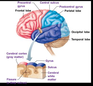
Regions of the Brain: Cerebrum
Surface Anatomy and Lobes
The cerebrum is divided into two hemispheres and further into lobes by prominent sulci and fissures.
Major Lobes: Frontal, parietal, occipital, and temporal.
Key Landmarks: Central sulcus (separates frontal and parietal lobes), parieto-occipital sulcus, lateral sulcus, and longitudinal fissure (separates hemispheres).
Gyri and Sulci: Ridges (gyri) and grooves (sulci) increase surface area for cortical processing.
Cerebral Cortex: Structure and Function
The cerebral cortex is a thin, highly convoluted layer of gray matter responsible for conscious thought, sensory perception, voluntary movement, communication, memory, and understanding.
Functional Areas:
Motor Areas: Control voluntary movement.
Sensory Areas: Conscious awareness of sensation.
Association Areas: Integrate diverse information for complex functions.
Each hemisphere controls the contralateral side of the body.
Conscious behavior involves the entire cortex.
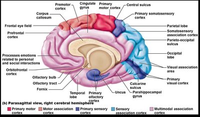
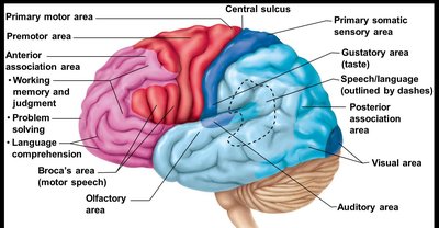
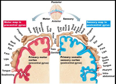
Specialized Areas of the Cerebrum
Distinct regions of the cortex are specialized for sensory and motor functions.
Primary Somatic Sensory Area: Located in the postcentral gyrus of the parietal lobe; receives sensory input from the body (pain, temperature, touch).
Primary Motor Area: Located in the precentral gyrus of the frontal lobe; initiates voluntary movements.
Motor and Sensory Homunculi: Spatial maps representing the distribution of motor and sensory control across the cortex.

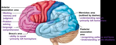
Association Areas and Special Senses
Association areas integrate sensory and motor information and are involved in higher cognitive functions.
Anterior Association Area (Prefrontal Cortex): Involved in judgment, reasoning, and planning.
Posterior Association Area: Integrates sensory input for understanding written and spoken language.
Broca's Area: Motor speech area (frontal lobe).
Wernicke's Area: Language comprehension (temporal lobe).
Special Senses: Visual (occipital lobe), auditory (temporal lobe), olfactory (temporal lobe), gustatory (parietal lobe).

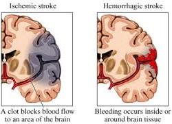
Cerebrovascular Accident (CVA) and Stroke
Pathophysiology and Effects
A stroke (CVA) results from a blocked or ruptured blood vessel in the brain, leading to tissue death and loss of function.
Ischemic Stroke: Caused by a clot blocking blood flow.
Hemorrhagic Stroke: Caused by bleeding into or around brain tissue.
Symptoms: Hemiplegia (paralysis on one side), aphasia (language impairment), and other neurological deficits.
Transient Ischemic Attack (TIA): Temporary restriction of blood flow; warning sign for stroke.
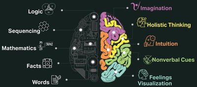
Lateralization of Cortical Function
Hemispheric Specialization
The two cerebral hemispheres are nearly identical but exhibit lateralization, meaning certain functions are more dominant in one hemisphere.
Left Hemisphere: Language, math, logic (dominant in ~90% of people).
Right Hemisphere: Visual-spatial skills, intuition, emotion, artistic and musical abilities.
Hemispheres communicate via the corpus callosum for functional integration.
Cerebral White and Gray Matter
White Matter: Fiber Tracts
Cerebral white matter consists of myelinated fibers that facilitate communication within the brain and between the brain and spinal cord.
Association Fibers: Connect regions within the same hemisphere.
Commissural Fibers: Connect corresponding regions of the two hemispheres (e.g., corpus callosum).
Projection Fibers: Connect the cortex with lower brain regions and the spinal cord.
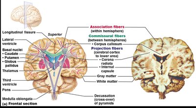
Basal Nuclei (Ganglia)
Basal nuclei are subcortical clusters of gray matter involved in regulating voluntary motor activity, cognition, and emotion.
Main Components: Caudate nucleus, putamen, globus pallidus (caudate + putamen = striatum).
Functions: Influence muscle movements, regulate movement intensity, filter inappropriate responses, and inhibit unnecessary movements.
Associated with the subthalamic nuclei and substantia nigra.

Regions of the Brain: Diencephalon
Major Components and Functions
The diencephalon is located atop the brain stem and is enclosed by the cerebral hemispheres. It consists of the thalamus, hypothalamus, and epithalamus.
Thalamus: Relay station for sensory impulses; directs information to the appropriate cortical areas.
Hypothalamus: Regulates autonomic functions, emotions, body temperature, hunger, thirst, sleep-wake cycles, and the endocrine system.
Epithalamus: Contains the pineal gland (endocrine) and choroid plexus (produces CSF).

Regions of the Brain: Brain Stem
Structure and Function
The brain stem connects the brain to the spinal cord and is essential for basic life functions.
Midbrain: Contains motor tracts, visual and auditory reflex centers, and nuclei for cranial nerves III and IV.
Pons: Relays information between the cerebrum and cerebellum; involved in breathing regulation; origin of cranial nerves V, VI, VII.
Medulla Oblongata: Controls autonomic functions such as heart rate, blood pressure, and respiration; origin of cranial nerves VIII–XII.
Regions of the Brain: Cerebellum
Structure and Function
The cerebellum coordinates voluntary movements, balance, and posture. It receives input from the cortex, brain stem, and sensory receptors.
Hemispheres: Connected by the vermis; each has anterior, posterior, and flocculonodular lobes.
Arbor Vitae: Tree-like arrangement of white matter.
Peduncles: Superior (to midbrain), middle (to pons), inferior (to medulla).
Role in Cognition: Involved in thinking, language, and emotion.
Functional Brain Systems
Limbic System
The limbic system is the emotional brain, involved in emotion, motivation, and memory formation.
Amygdaloid Body: Recognizes emotions and assesses danger.
Cingulate Gyrus: Expresses emotions via gestures and resolves mental conflict.
Hippocampus: Essential for memory consolidation and retrieval.
Reticular Formation
The reticular formation is a network of neurons running through the brain stem, crucial for arousal and consciousness.
Reticular Activating System (RAS): Maintains alertness and filters sensory input.
Motor Function: Controls coarse limb movements and regulates visceral motor functions.
Protection of the Central Nervous System
Five Protective Mechanisms
The CNS is protected by multiple anatomical and physiological barriers.
Scalp and Skin: First line of defense.
Skull and Vertebral Column: Bony encasement for the brain and spinal cord.
Meninges: Three connective tissue membranes (dura mater, arachnoid mater, pia mater) that cover the CNS.
Cerebrospinal Fluid (CSF): Cushions and nourishes the brain and spinal cord.
Blood-Brain Barrier: Selectively restricts passage of substances from the blood into the CNS.
Cerebrospinal Fluid (CSF)
CSF is a clear, colorless fluid produced by the choroid plexuses in the ventricles. It provides buoyancy, protection, and chemical stability for the CNS.
Composition: Similar to plasma but with less protein and different ion concentrations.
Functions: Reduces brain weight, protects from trauma, nourishes, and removes waste.
Flow: Circulates through ventricles, subarachnoid space, and central canal; reabsorbed by arachnoid villi.
Blood-Brain Barrier
The blood-brain barrier is formed by endothelial cells with tight junctions, astrocyte feet, and a thick basal lamina. It allows selective passage of substances to protect the brain from toxins and pathogens.
Permitted: Nutrients, fat-soluble substances, some gases.
Restricted: Metabolic wastes, proteins, toxins, most drugs.
Absent: In areas where blood monitoring is necessary (e.g., hypothalamus).
Spinal Cord: Gross Anatomy and Protection
Structure and Function
The spinal cord extends from the foramen magnum to the first or second lumbar vertebra and is protected by bone, meninges, and CSF.
Conus Medullaris: Tapered end of the spinal cord.
Filum Terminale: Fibrous extension anchoring the cord to the coccyx.
Denticulate Ligaments: Extensions of pia mater securing the cord laterally.
Spinal Nerves: 31 pairs, part of the peripheral nervous system (PNS).
Cauda Equina: Bundle of nerve roots at the inferior end of the vertebral canal.
Internal Structure
Gray Matter: Central, butterfly-shaped region containing neuron cell bodies; divided into dorsal (sensory) and ventral (motor) horns.
White Matter: Surrounds gray matter; contains ascending (sensory) and descending (motor) tracts.
Central Canal: Contains CSF.
Ascending and Descending Pathways
Ascending (Sensory) Pathways:
Dorsal columns: Discriminatory touch and vibration.
Spinocerebellar tracts: Proprioceptive information to cerebellum.
Spinothalamic tracts: Pain, temperature, crude touch, and pressure.
Descending (Motor) Pathways:
Corticospinal tracts: Voluntary motor control.
Reticulospinal, vestibulospinal, and tectospinal tracts: Muscle tone, balance, and reflexive movements.
Disorders of the Central Nervous System
Major Disorders
Autism: Developmental disorder affecting communication and social interaction.
Huntington's Disease: Hereditary, fatal disorder with degeneration of basal nuclei and cerebral cortex, leading to motor and cognitive decline.
Anencephaly: Failure of the cerebrum to develop.
Spina Bifida: Incomplete formation of vertebrae, exposing the spinal cord.
Meningitis: Inflammation of the meninges due to infection.
