 Back
BackChapter 11: Introduction to the Nervous System and Nervous Tissue - Study Notes
Study Guide - Smart Notes
 Tailored notes based on your materials, expanded with key definitions, examples, and context.
Introduction to the Nervous System
Overview of the Nervous System
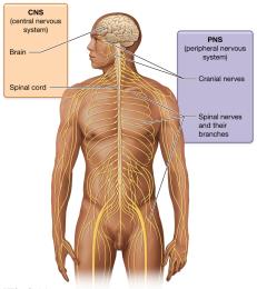
The nervous system is a complex network that controls perception, voluntary movement, consciousness, personality, learning, and memory. It works closely with the endocrine system to maintain homeostasis, regulating variables such as respiratory rate, blood pressure, body temperature, sleep/wake cycle, and blood pH.
Central Nervous System (CNS): Consists of the brain and spinal cord.
Peripheral Nervous System (PNS): Composed of cranial nerves, spinal nerves, and their branches.
Functional Divisions of the Nervous System
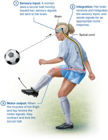
The nervous system is divided functionally into sensory, integrative, and motor divisions, each responsible for distinct physiological roles.
Sensory (Afferent) Division: Gathers information from internal and external environments via sensory receptors.
Integrative Functions: Analyze and interpret sensory stimuli, determining appropriate responses (primarily in the CNS).
Motor (Efferent) Division: Executes actions in response to integration, targeting effectors such as muscles and glands.
Structural and Functional Summary
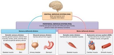
The nervous system is organized into structural and functional divisions, with specialized sensory and motor pathways.
Somatic Sensory Division: Signals from skeletal muscles, bones, joints, skin, and special senses.
Visceral Sensory Division: Signals from internal organs (viscera).
Somatic Motor Division: Voluntary control of skeletal muscles.
Autonomic Nervous System (ANS): Involuntary control of glands, smooth muscle, and cardiac muscle.
Nervous Tissue
Cellular Composition of Nervous Tissue
Nervous tissue is composed of cells (about 80% of its volume) and extracellular matrix (about 20%). The two main cell types are neurons and neuroglial cells.
Neurons: Excitable cells responsible for transmitting electrical signals.
Neuroglial Cells: Supportive cells that maintain the environment, protect neurons, and assist in their functions.
Neurons
Structure and Function of Neurons
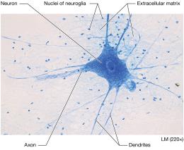
Neurons are specialized for sending and receiving signals via action potentials. They are generally long-lived and amitotic, with a central cell body, dendrites, and a single axon.
Cell Body (Soma): Contains most organelles, including Nissl bodies (clusters of ribosomes and rough ER), Golgi apparatus, nucleoli, and mitochondria.
Dendrites: Short, highly branched processes that receive input and transmit electrical signals toward the cell body.
Axon: Long process that carries electrical signals away from the cell body; includes axon hillock, axon collaterals, telodendria, and axon terminals.
Functional Regions of Neurons
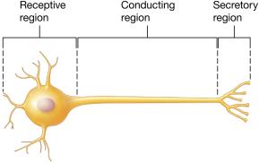
Each neuron has three main functional regions:
Receptive Region: Dendrites and cell body receive signals.
Conducting Region: Axon transmits the signal.
Secretory Region: Axon terminals release neurotransmitters to target cells.
Classification of Neurons
Neurons are classified structurally and functionally:
Multipolar Neurons: One axon, multiple dendrites; most common in CNS.
Bipolar Neurons: One axon, one dendrite; found in special sense organs.
Pseudounipolar Neurons: Single process splits into two axons; sensory neurons for touch, pain, and vibration.
Sensory (Afferent) Neurons: Carry signals toward CNS.
Interneurons: Relay messages within CNS.
Motor (Efferent) Neurons: Carry signals away from CNS to effectors.
Groups of Neuron Cell Bodies and Axons
Nuclei: Clusters of cell bodies in CNS.
Ganglia: Clusters of cell bodies in PNS.
Tracts: Bundles of axons in CNS.
Nerves: Bundles of axons in PNS.
Neuroglia (Neuroglial Cells)
Types and Functions of Neuroglia
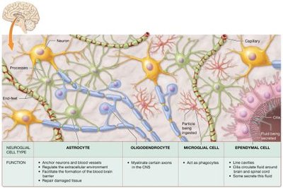
Neuroglia support neurons, maintain their environment, and protect them. They can undergo mitosis and fill gaps when neurons die. There are six types: four in the CNS and two in the PNS.
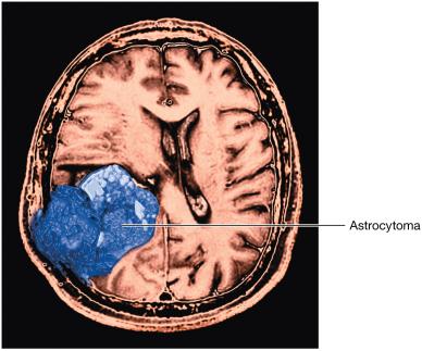
Astrocytes: Anchor neurons and blood vessels, regulate extracellular environment, assist in blood-brain barrier formation, and repair damaged tissue.
Oligodendrocytes: Form myelin sheaths in CNS, increasing speed of impulse conduction.
Microglia: Act as phagocytes, ingesting pathogens and debris, and stimulate inflammation.
Ependymal Cells: Circulate and monitor cerebrospinal fluid in CNS.
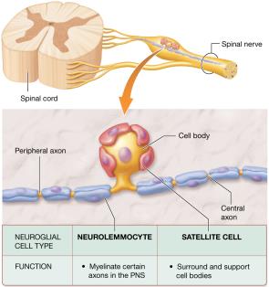
Neurolemmocytes (Schwann Cells): Myelinate axons in PNS and assist in axon repair.
Satellite Cells: Surround and support cell bodies in PNS, regulating the extracellular environment.
The Myelin Sheath
Structure and Function of Myelin
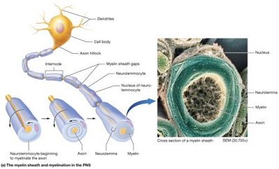
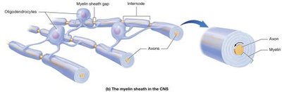
The myelin sheath is formed by neurolemmocytes in the PNS and oligodendrocytes in the CNS. It consists of repeating layers of plasma membrane, acting as an insulator and increasing the speed of action potential conduction.
Myelinated Axons: Conduct action potentials 15–150 times faster than unmyelinated axons.
Internodes: Segments of axon covered by myelin.
Nodes of Ranvier: Gaps between internodes where ion channels are concentrated.
Unmyelinated Axons
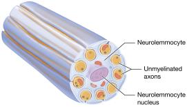
Short axons are typically unmyelinated. In the PNS, unmyelinated axons are associated with neurolemmocytes. In the CNS, myelinated areas appear as white matter, while unmyelinated areas are gray matter.
Regeneration of Nervous Tissue
Repair Mechanisms
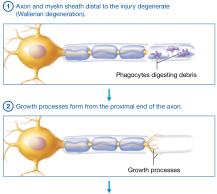
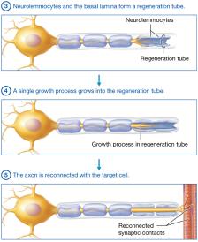
Regeneration is limited in the CNS but possible in the PNS if the cell body remains intact. The process involves degeneration, growth, formation of a regeneration tube, and reconnection with the target cell.
Wallerian Degeneration: Axon and myelin distal to injury degenerate; phagocytes digest debris.
Growth Processes: Form from the proximal end of the axon.
Regeneration Tube: Formed by neurolemmocytes and basal lamina.
Reconnection: Axon reconnects with the target cell.
Electrophysiology of Neurons
Resting Membrane Potential
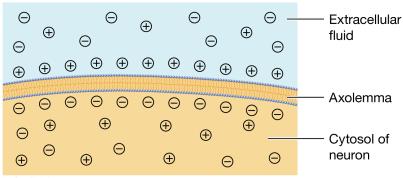
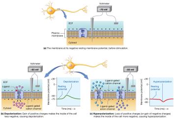
Neurons are excitable and respond to stimuli by generating electrical changes across their plasma membranes. The resting membrane potential is typically -70 mV, maintained by ion gradients and channels.
Polarization: The membrane potential is negative at rest.
Depolarization: Membrane potential becomes less negative.
Repolarization: Return to resting potential.
Hyperpolarization: Membrane potential becomes more negative than at rest.
Ion Channels and Pumps
Ions cross the plasma membrane via leak channels, ligand-gated channels, voltage-gated channels, and mechanically gated channels. The sodium-potassium pump maintains ion gradients by moving 2 K+ into the cell and 3 Na+ out.
Action Potentials
Generation and Propagation
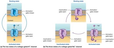
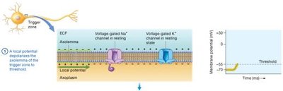
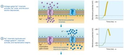
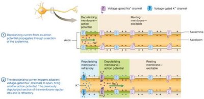
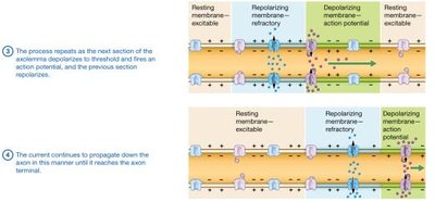
Action potentials are rapid, uniform depolarizations and repolarizations that travel along axons. They are generated at the trigger zone and involve voltage-gated sodium and potassium channels.
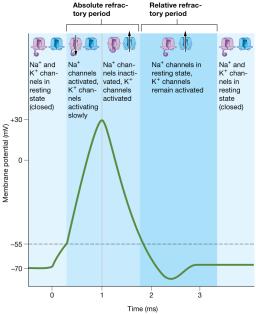
Depolarization: Sodium channels open, Na+ enters, membrane potential rises to +30 mV.
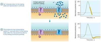
Repolarization: Sodium channels inactivate, potassium channels open, K+ exits.
Hyperpolarization: Membrane potential may become more negative before returning to rest.
Refractory Period
The refractory period is the time after an action potential when the neuron cannot fire another action potential. It includes the absolute refractory period (no stimulus can trigger another action potential) and the relative refractory period (a stronger stimulus can trigger another action potential).
Propagation of Action Potentials
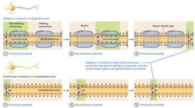
Action potentials are self-propagating and travel in one direction along the axon. Conduction speed depends on axon diameter and myelination.
Continuous Conduction: Occurs in unmyelinated axons; each section must be depolarized.
Saltatory Conduction: Occurs in myelinated axons; action potentials jump between nodes of Ranvier.
Neuronal Synapses
Types of Synapses
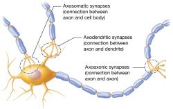
Synapses are junctions where neurons communicate with target cells. Neuronal synapses can be axodendritic, axosomatic, or axoaxonic.
Electrical and Chemical Synapses
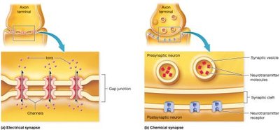
Electrical synapses involve gap junctions and allow rapid, bidirectional transmission. Chemical synapses use neurotransmitters and are unidirectional, with a synaptic delay.
Events at a Chemical Synapse
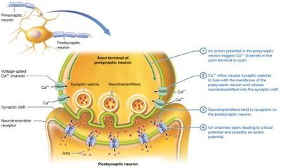
Action potential triggers Ca2+ influx in presynaptic neuron.
Synaptic vesicles release neurotransmitters into the synaptic cleft.
Neurotransmitters bind to postsynaptic receptors, opening ion channels and generating local potentials.
Postsynaptic Potentials
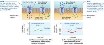
Excitatory Postsynaptic Potential (EPSP): Depolarizes the postsynaptic membrane.
Inhibitory Postsynaptic Potential (IPSP): Hyperpolarizes the postsynaptic membrane.
Summation and Neural Integration
Temporal Summation: Rapid, repeated EPSPs from a single presynaptic neuron.
Spatial Summation: Simultaneous EPSPs from multiple presynaptic neurons.
Channels and Pumps in Neurons
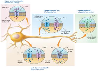
Ligand-Gated Channels: Found on dendrites and cell body.
Voltage-Gated Na+ and K+ Channels: Found on axon.
Voltage-Gated Ca2+ Channels: Found at axon terminal.
Leak Channels and Na+/K+ Pump: Found throughout the neuron.
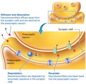
Termination of Synaptic Transmission
Diffusion and Absorption: Neurotransmitters diffuse away and are returned to the presynaptic neuron.
Degradation: Enzymatic breakdown in the synaptic cleft.
Reuptake: Neurotransmitters are taken back into the presynaptic neuron.
Neurotransmitter Receptors and Major Neurotransmitters
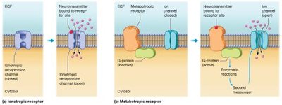
Types of Neurotransmitter Receptors
Ionotropic Receptors: Part of ligand-gated ion channels; rapid, short-lived effects.
Metabotropic Receptors: Linked to G-proteins and second messengers; slower, longer-lasting effects.
Major Neurotransmitters
Acetylcholine (ACh): Excitatory at neuromuscular junctions; broken down by acetylcholinesterase.
Biogenic Amines: Includes norepinephrine, epinephrine, dopamine, serotonin, and histamine; diverse functions in CNS and PNS.
Amino Acid Neurotransmitters: Glutamate (excitatory), GABA and glycine (inhibitory).
Neuropeptides: Substance P (pain), opioids (pain relief), neuropeptide Y (feeding behavior).
Neuronal Pools and Circuits
Neuronal Pools
Groups of interneurons in the CNS that perform common functions, defined by synaptic connections. Input neurons initiate signals, which may generate EPSPs or IPSPs.
Neuronal Circuits
Diverging Circuit: One input neuron branches to multiple postsynaptic neurons.
Converging Circuit: Multiple input neurons converge onto a single postsynaptic neuron.
Inhibitory Mechanisms: Prevent excessive excitation via neuromodulators and synaptic fatigue.
Epileptic Seizures
Epilepsy is characterized by recurrent episodes of abnormal electrical activity in the brain, resulting from a sudden burst of excitatory activity that overwhelms inhibitory circuits. Symptoms include sensory disturbances, loss of consciousness, and jerking movements. Treatment aims to prevent seizures and restore inhibitory function.
