 Back
BackChapter 11: The Nervous System & Nervous Tissue – Structured Study Notes
Study Guide - Smart Notes
 Tailored notes based on your materials, expanded with key definitions, examples, and context.
Chapter 11 – The Nervous System & Nervous Tissue
The Nervous System: Overview
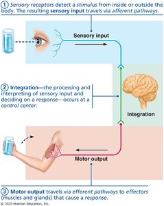
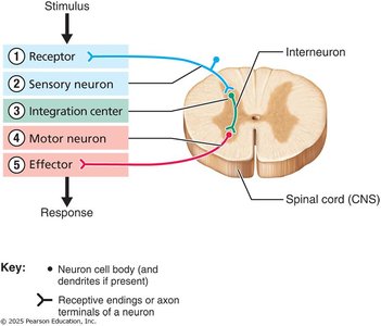
The nervous system is the primary controlling and communicating system of the body, utilizing rapid electrical and chemical signals to coordinate responses. It performs three main functions: sensory input, integration, and motor output.
Sensory Input: Detects internal and external changes via sensory receptors. Information is sent to the central nervous system (CNS) through afferent pathways.
Integration: Processes and interprets sensory input, deciding on an appropriate response. This occurs in the CNS, primarily the brain or spinal cord.
Motor Output: Activates effector organs (muscles or glands) to produce a response, sent from the CNS via efferent pathways.
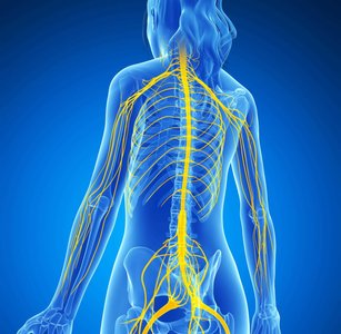
Levels of Organization in the Nervous System
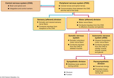
The nervous system is organized into the central nervous system (CNS) and peripheral nervous system (PNS), each with distinct structures and functions.
CNS: Composed of the brain and spinal cord; serves as the integration and control center.
PNS: Includes all nervous structures outside the CNS (cranial nerves, spinal nerves, ganglia, and branches).
PNS Functional Divisions:
Sensory (afferent) division: Sends signals toward the CNS.
Motor (efferent) division: Sends signals away from the CNS.
Motor Division Subdivisions:
Somatic nervous system: Controls voluntary movements by connecting CNS to skeletal muscles.
Autonomic nervous system (ANS): Controls involuntary functions (cardiac, smooth muscle, glands).
Sympathetic division: Mobilizes body systems during activity (fight or flight).
Parasympathetic division: Promotes maintenance functions during rest (rest and digest).
Nervous Tissue Histology
Nervous tissue is highly cellular, with minimal extracellular space. It consists of two principal cell types: neuroglia and neurons.
Neuroglia: Support and protect neurons. Types include astrocytes, microglial cells, ependymal cells, oligodendrocytes (CNS), satellite cells, and Schwann cells (PNS).
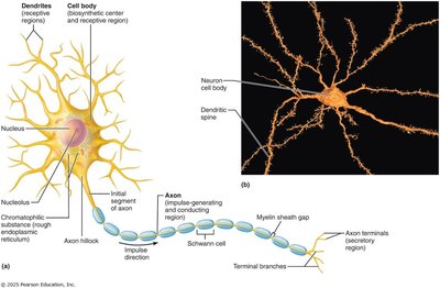
Neurons: Excitable cells that transmit electrical signals. They have a cell body (soma) and one or more processes (dendrites and axons).
Neurons: Structure and Function
Neurons are the structural and functional units of the nervous system, specialized for impulse conduction. They exhibit extreme longevity, are amitotic, and have a high metabolic rate.
Cell Body (Soma): Biosynthetic center, synthesizes proteins, membranes, and chemicals.
Processes:
Dendrites: Receive stimuli and carry information toward the cell body as graded potentials.
Axons: Carry signals away from the cell body, connect with other neurons or effectors, and transmit action potentials.
Axons and Myelin Sheaths
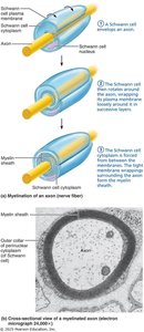
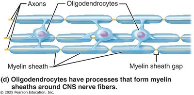
Axons are the conducting regions of neurons, generating and transmitting nerve impulses. Myelin sheaths, composed of a protein-lipid substance, surround most long or large-diameter axons, providing protection and increasing transmission speed.
Myelin in CNS: Formed by oligodendrocytes; one cell forms multiple sheaths.
Myelin in PNS: Formed by Schwann cells; multiple cells form one sheath. Schwann cells also aid in nerve regeneration.
Neuron Classification
Neurons are classified structurally by the number of processes and functionally by the direction of impulse transmission.
Structural Classification:
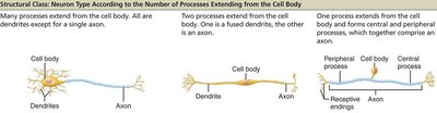
Multipolar: Most common; major CNS neuron (one axon, multiple dendrites).
Bipolar: Rare; found in retina and olfactory mucosa (one axon, one dendrite).
Unipolar: One short process divides into two branches (peripheral and central processes).
Functional Classification:
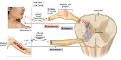
Sensory (afferent): Carries impulses toward CNS; mostly unipolar.
Motor (efferent): Carries impulses away from CNS to effectors; mostly multipolar.
Interneurons: Transfer impulses within CNS; multipolar; majority of neurons.
Resting Membrane Potential
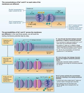
The resting membrane potential is an electrochemical difference across the cell membrane, resulting from ion concentration gradients and selective permeability.
Inside of cell: Negatively charged relative to outside due to K+ leakage channels and Na+ leakage channels.
Na/K Pump: Maintains gradients by exporting 3 Na+ for every 2 K+ imported, requiring ATP.
Membrane Potentials: Graded and Action Potentials
Neurons use two types of membrane potentials as signals: graded potentials (short-distance, lose strength) and action potentials (long-distance, do not lose strength).
Graded Potentials: Incoming signals, lose strength with distance.
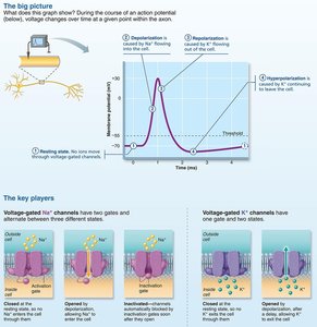
Action Potentials: Long-distance signals, principal means of neural communication.
Ion Channels in Neurons
Neurons possess various ion channels that regulate membrane potential and signal transmission.
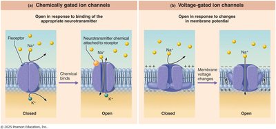
Leakage Channels: Always open, allow passive ion movement.
Gated Channels:
Chemically gated: Open with neurotransmitter binding.
Voltage-gated: Open/close in response to membrane potential changes.
Mechanically gated: Open/close in response to physical deformation.
The Synapse
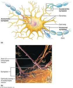
Synapses are junctions that mediate information transfer between neurons. They can be chemical (most common) or electrical (rare).
Chemical Synapses: Neurotransmitters bridge the gap between presynaptic and postsynaptic neurons.
Electrical Synapses: Neurons are electrically coupled via gap junctions.
Neurotransmitters
Neurotransmitters are chemical messengers used by neurons to communicate. Over 50 have been identified, classified by structure and function.
Acetylcholine (ACh): First identified, released at neuromuscular junctions and by some CNS/ANS neurons. Can stimulate or inhibit responses.
Neural Processing: Serial and Parallel
Neural processing can occur in serial or parallel pathways.
Serial Processing: Information travels in a single pathway, producing predictable responses (e.g., spinal reflexes).
Parallel Processing: Input travels along multiple pathways simultaneously, enabling complex responses and higher-level mental functions.
Summary Table: Nervous System Organization
Division | Main Structures | Function |
|---|---|---|
CNS | Brain, Spinal Cord | Integration, control center |
PNS | Cranial nerves, spinal nerves, ganglia | Communication between CNS and body |
Sensory (afferent) | Sensory receptors, nerve fibers | Transmit signals to CNS |
Motor (efferent) | Motor nerve fibers | Transmit signals from CNS to effectors |
Somatic | Skeletal muscles | Voluntary movement |
Autonomic | Cardiac, smooth muscle, glands | Involuntary functions |
Sympathetic | Various organs | Fight or flight |
Parasympathetic | Various organs | Rest and digest |
Key Equations
Resting Membrane Potential:
Na/K Pump:
Additional info:
Neurons are amitotic, meaning they do not divide after development.
Schwann cells in the PNS facilitate regeneration of damaged axons, unlike oligodendrocytes in the CNS.
