 Back
Chapter 12: Nervous System and Nervous Tissue – Structured Study Notes
Study Guide - Smart Notes
 Tailored notes based on your materials, expanded with key definitions, examples, and context.
Nervous System and Nervous Tissue
Overview and Functions of the Nervous System
The nervous system is responsible for monitoring internal and external changes, processing information, and responding to stimuli. It is essential for sensory perception, integration, and motor output.
Sensory Input: Detection of changes (stimuli) via sensory receptors.
Integration: Processing and interpretation of sensory input in the central nervous system (CNS).
Motor Output: Response to stimuli by activating effectors such as muscles and glands.
Afferent: Signals traveling toward the CNS.
Efferent: Signals traveling away from the CNS.
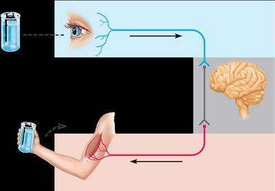
Structural Divisions of the Nervous System
The nervous system is divided into two main anatomical divisions:
Central Nervous System (CNS): Consists of the brain and spinal cord, located in the cranium and vertebral canal.
Peripheral Nervous System (PNS): Includes cranial nerves, spinal nerves, and ganglia. Cranial nerves carry signals to and from the brain; spinal nerves carry signals to and from the spinal cord; ganglia are clusters of neuronal cell bodies.
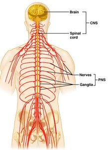
Functional Divisions in the PNS
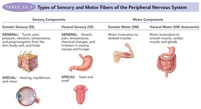
Sensory (Afferent) Division: Transmits sensory information to the CNS.
Motor (Efferent) Division: Transmits motor commands from the CNS to effectors.
Somatic Nervous System: Controls voluntary movements of skeletal muscles.
Autonomic Nervous System: Controls involuntary functions (cardiac/smooth muscle, glands). Includes sympathetic (fight or flight) and parasympathetic (rest and digest) divisions.
Cells of Nervous Tissue
Nervous tissue consists of two main cell types:
Neurons: Excitable cells that transmit electrical signals.
Neuroglia (Glial Cells): Non-excitable supportive cells that maintain the environment for neurons.
Neurons: Structure and Function
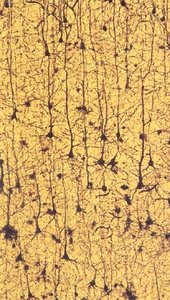
Neurons are the fundamental units of function in the CNS, with unique properties and specialized structures.
Cell Body (Soma): Contains the nucleus, nucleolus, and organelles; biosynthetic center of the neuron.
Dendrites: Branching processes that receive signals and conduct them toward the cell body.
Axon: Single, long process that conducts impulses away from the cell body; may be myelinated.
Synaptic Terminals: Endings of the axon that contain synaptic vesicles filled with neurotransmitters.
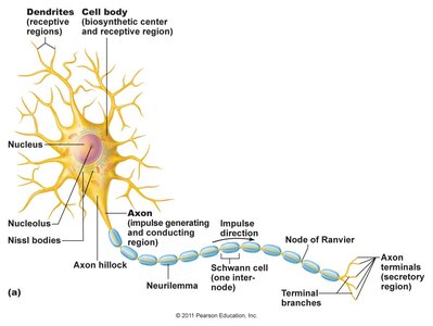
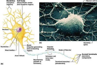
Neuron Cell Body (Soma)
Diameter: 5–140 μm
Single nucleus with large nucleolus
Contains Nissl bodies (clusters of rough ER), mitochondria, lysosomes, Golgi apparatus
Renews cell membranes and cytosolic proteins
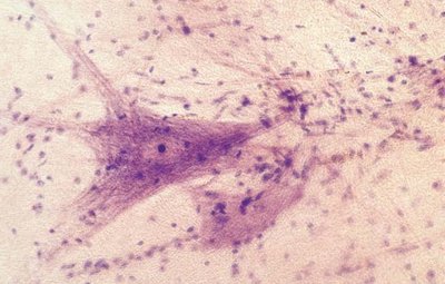
Dendrites
Numerous branching processes
Receptive sites for incoming signals
Increase surface area for signal reception
Conduct electrical signals toward the cell body
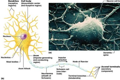
Axon
One per neuron, uniform diameter
Ends in multiple terminals
May have collateral branches
Axon terminals contain synaptic vesicles
Impulse generation at initial segment; conduction away from cell body
Often myelinated
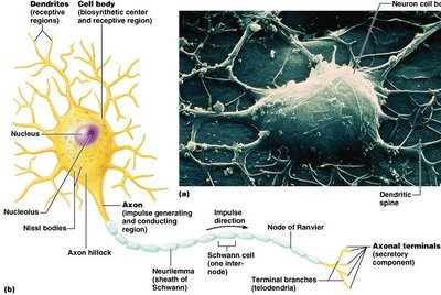
Synapses: Structure and Function
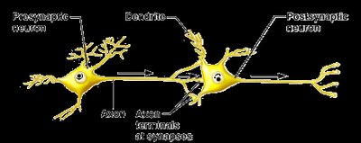
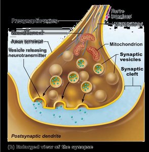
Synapses are specialized junctions where neurons communicate with other neurons or effectors. Most synapses are chemical, involving neurotransmitter release.
Presynaptic Neuron: Releases neurotransmitter into the synaptic cleft.
Synaptic Cleft: Space between presynaptic and postsynaptic membranes.
Postsynaptic Neuron: Contains receptors for neurotransmitters.
Neurotransmitters can be excitatory or inhibitory.
Classification of Neurons
Structural Classification
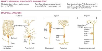
Neurons are classified based on the number and arrangement of their processes:
Multipolar: Multiple dendrites, one axon; most abundant (>99% of all neurons).
Bipolar: One dendrite, one axon; rare, found in special sensory organs.
Unipolar: Single process; most sensory neurons.
Functional Classification
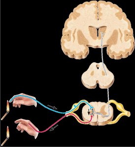
Sensory Neurons: Transmit sensory information toward the CNS.
Motor Neurons: Transmit motor commands from the CNS to effectors.
Interneurons: Confined to the CNS; integrate and process information.
Neuroglial Cells (Glia)
Glial cells support and maintain the neural environment. They are mitotically active and can give rise to brain tumors.
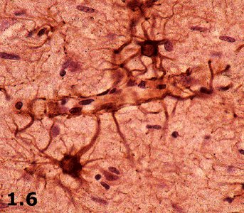
Astrocytes: Most numerous; maintain ionic homeostasis, support blood-brain barrier, promote neuronal growth.
Oligodendrocytes: Myelinate axons in the CNS; one cell can myelinate several axons.
Schwann Cells: Myelinate axons in the PNS; one cell myelinates one axon.
Microglia: Macrophages of the CNS; remove debris and act as immune cells.
Ependymal Cells: Form epithelial lining of brain ventricles; produce and circulate cerebrospinal fluid (CSF).
Myelin Sheath
Myelin sheaths are concentric layers of plasma membrane produced by glial cells, wrapping around axons.
Protects axons and increases speed of impulse transmission.
Electrically insulates nerve fibers from one another.
Schwann Cells: Form myelin sheaths in the PNS (one cell per axon).
Oligodendrocytes: Form myelin sheaths in the CNS (one cell for several axons).
Nodes of Ranvier: Gaps in the myelin sheath where impulse conduction is facilitated.
Gray Matter vs. White Matter in the CNS
Gray Matter: Contains neuronal cell bodies, dendrites, and axon terminals; site of synapses. In the spinal cord, it forms an H-shaped internal region; in the brain, it is found in the cortex and deep areas.
White Matter: Composed of myelinated axons; organized in tracts that connect different regions.
Nerves: Structure and Components
Nerves are organs of the PNS, consisting of bundles of nerve fibers (axons), Schwann cells, blood vessels, and connective tissue layers.
Endoneurium: Surrounds a single axon.
Perineurium: Surrounds a bundle of axons (fascicle).
Epineurium: Surrounds the whole nerve.
Most nerves contain both sensory and motor fibers, which can be myelinated or unmyelinated.
Summary Table: Types of Sensory and Motor Fibers in the PNS
Type | Sensory Components | Motor Components |
|---|---|---|
Somatic | Touch, pain, pressure, vibration, temperature, proprioception (skin, body wall, limbs) | Motor innervation to skeletal muscles |
Visceral | Stretch, pain, temperature, chemical changes, irritation (internal organs) | Motor innervation to smooth muscle, cardiac muscle, glands |
Special | Hearing, equilibrium, vision, taste, smell | --- |
Key Equations and Concepts
Resting Membrane Potential:
Action Potential Propagation:
Additional info:
Interneurons constitute >99% of all neurons in the CNS and are essential for complex processing and integration.
Loss of myelination (e.g., in multiple sclerosis) severely impairs nervous system function.
