 Back
BackChapter 12: The Central Nervous System – Structure, Function, and Protection
Study Guide - Smart Notes
 Tailored notes based on your materials, expanded with key definitions, examples, and context.
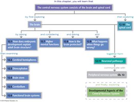
Chapter Overview: The Central Nervous System
The central nervous system (CNS) is composed of the brain and spinal cord, serving as the primary control center for the body. This chapter explores the major anatomical structures, functional regions, protective coverings, and developmental aspects of the CNS.
Latin and Greek Terminology in Neuroanatomy
Key Terms and Etymology
Understanding the roots of neuroanatomical terms aids in mastering the vocabulary of the CNS.
Cephalo-: Head
Cerebral: Pertaining to the brain
Encephalo-: In the head
Myelo-: Marrow, spinal cord (related to myelin)
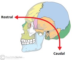
Rostral: Toward the beak (front)
Caudal: Toward the tail (back)
Ventricle: Little belly (cavity in the brain)

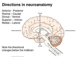
Directional Terms in Neuroanatomy
Understanding Anatomical Directions
Directional terms are essential for describing locations and orientations within the CNS.
Anterior–Posterior: Front to back
Rostral–Caudal: Toward the nose/beak to toward the tail
Dorsal–Ventral: Back to belly
Superior–Inferior: Above to below
Medial–Lateral: Toward the midline to away from the midline
CNS Major Structures
Overview from Rostral to Caudal
The CNS is organized from the most rostral (anterior) to the most caudal (posterior) structures:
Cerebrum (2 cerebral hemispheres)
Diencephalon (thalamus, hypothalamus, etc.)
Cerebellum
Brain Stem (midbrain, pons, medulla oblongata)
Spinal Cord
Cerebral Hemispheres
Structure and Communication
The cerebrum consists of two hemispheres separated by the longitudinal fissure and connected by the corpus callosum. The wrinkled surface increases cortical area for higher processing.
Gyri: Elevated ridges ("hills")
Sulci: Shallow grooves ("valleys")
Fissures: Deep grooves
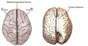
Corpus Callosum
The corpus callosum is a thick band of nerve fibers that enables communication between the right and left hemispheres.
Hemispheric Lateralization
Each hemisphere has specialized functions, though they work together:
Left Hemisphere: Language, logic, math
Right Hemisphere: Art, emotion, intuition, spatial skills
Some individuals may have reversed or shared dominance.
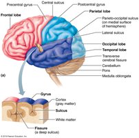
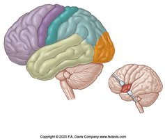
Lobes of the Cerebrum
Each hemisphere is divided into five lobes, separated by sulci:
Frontal Lobe: Voluntary movement, executive functions
Parietal Lobe: Somatosensation (body sensation)
Temporal Lobe: Hearing, smell, memory
Occipital Lobe: Vision
Insula: Taste, visceral sensation (internal)
Functional Areas of the Cerebral Cortex
Specific regions of the cortex are responsible for distinct functions:
Broca’s Area: Motor speech (usually left hemisphere)
Wernicke’s Area: Language comprehension
Precentral Gyrus: Primary motor cortex
Postcentral Gyrus: Primary somatosensory cortex
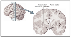
Gray and White Matter
The cerebrum is organized into layers:
Cerebral Cortex: Superficial gray matter (neuron cell bodies, dendrites)
White Matter: Deeper myelinated axon tracts
Basal Nuclei: Deep gray matter involved in movement control
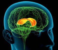
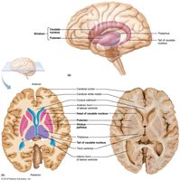
Basal Nuclei (Basal Ganglia)
Basal nuclei include the caudate nucleus, putamen, and globus pallidus. They regulate movement initiation, intensity, and termination. Disorders include Parkinson’s and Huntington’s diseases.
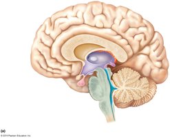
Diencephalon
Major Components and Functions
The diencephalon is located deep within the brain and includes:
Thalamus: Relay station for sensory information to the cortex
Hypothalamus: Homeostasis, autonomic and endocrine control, regulates temperature, hunger, thirst, sleep-wake cycles, and links to the pituitary gland
Epithalamus: Contains the pineal gland, which produces melatonin for sleep regulation
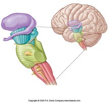
Brain Stem
Structure and Basic Life Functions
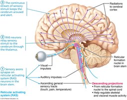
The brain stem consists of the midbrain, pons, and medulla oblongata. It controls vital functions such as breathing and heart rate, houses cranial nerve nuclei, and contains the reticular activating system (RAS) for consciousness and alertness.
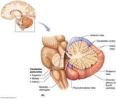
Cerebellum
Coordination and Balance
The cerebellum, separated from the cerebrum by the transverse cerebral fissure, has two hemispheres and a highly folded surface. It fine-tunes motor activity, balance, and coordination. The arbor vitae is the tree-like arrangement of white matter within the cerebellum.
Functional Brain Systems
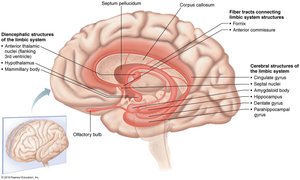
Limbic System and Reticular Activating System (RAS)
Functional systems span multiple brain regions:
Limbic System: Emotional and visceral responses, includes structures in the cerebrum and diencephalon
RAS: Maintains alertness and consciousness, spans the brain stem and influences the cerebrum
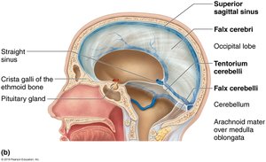
Protection of the CNS
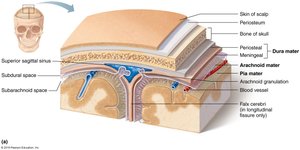
Meninges
The CNS is protected by three connective tissue membranes called meninges:
Dura Mater: Tough, outermost layer with periosteal and meningeal layers
Arachnoid Mater: Middle, web-like layer filled with cerebrospinal fluid (CSF)
Pia Mater: Delicate, vascular inner layer adhering to the brain and spinal cord
Spaces:
Subdural Space: Between dura and arachnoid mater
Subarachnoid Space: Between arachnoid and pia mater, contains CSF
Blood Supply and Blood Brain Barrier (BBB)
The brain receives blood from the vertebral and internal carotid arteries, which form the Circle of Willis. The BBB is a selective barrier formed by endothelial tight junctions, astrocytes, and pericytes, protecting the CNS from harmful substances but also limiting drug delivery.
Cerebrospinal Fluid (CSF)
CSF is produced by the choroid plexus in the ventricles, circulates through the ventricular system and subarachnoid space, and is reabsorbed into the blood at arachnoid granulations. CSF cushions the CNS and removes waste.
Developmental Aspects of the CNS
Neural Tube Formation and Defects
The CNS develops from the neural tube, which forms early in embryonic development. Failure of the neural tube to close can result in defects such as anencephaly (rostral end) or spina bifida (caudal end). Folic acid supplementation before and during early pregnancy greatly reduces the risk of neural tube defects.
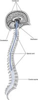
The Spinal Cord
Structure and Function
The spinal cord extends from the foramen magnum to the level of L1/L2. It transmits sensory information to the brain and motor commands from the brain, and mediates reflexes.
Cervical and Lumbar Enlargements: For limb innervation
Conus Medullaris: Tapered end of the spinal cord
Cauda Equina: Bundle of nerve roots below the spinal cord
Filum Terminale: Fibrous extension anchoring the cord to the coccyx
Cross-Sectional Anatomy
Gray Matter: H-shaped, contains neuron cell bodies; divided into dorsal (sensory), ventral (motor), and lateral (autonomic) horns
White Matter: Myelinated axon tracts; divided into dorsal (sensory), lateral (mixed), and ventral (motor) columns
Spinal Meninges
The spinal cord is covered by the same three meningeal layers as the brain. The pia mater forms denticulate ligaments and the filum terminale for anchoring.
Neuronal Pathways
Descending (Motor) Pathways
Motor commands travel from the brain to the body via two main pathways:
Direct (Pyramidal) Pathways: Two-neuron chain (upper and lower motor neurons) for voluntary movement
Indirect (Extrapyramidal) Pathways: Multineuronal, control balance, posture, and coarse movements
Ascending (Sensory) Pathways
Sensory information ascends to the brain via three-neuron chains:
Dorsal Columns: Touch and proprioception (cross in medulla)
Spinothalamic Tracts: Pain and temperature (cross in spinal cord)
Spinocerebellar Tracts: Subconscious proprioception
Decussation
Most pathways cross (decussate) to the opposite side of the CNS, explaining why brain injuries affect the contralateral side of the body.
Clinical Note: Brown-Sequard Syndrome
Hemisection of the spinal cord causes loss of touch/proprioception on the same side and pain/temperature on the opposite side below the lesion.
Summary Table: Major CNS Structures and Functions
Structure | Main Function(s) |
|---|---|
Cerebrum | Higher cognitive functions, voluntary movement, sensory perception |
Diencephalon | Sensory relay, homeostasis, endocrine regulation |
Brain Stem | Basic life functions, cranial nerves, consciousness |
Cerebellum | Coordination, balance, fine motor control |
Spinal Cord | Sensory/motor relay, reflexes |
Additional info: This guide integrates foundational concepts from OpenStax, Marieb & Hoehn, and F.A. Davis, with expanded academic context for clarity and exam preparation.
