 Back
BackChapter 12: The Nervous System and Nervous Tissue – Structured Study Notes
Study Guide - Smart Notes
 Tailored notes based on your materials, expanded with key definitions, examples, and context.
The Nervous System and Nervous Tissue
Overview of the Nervous System
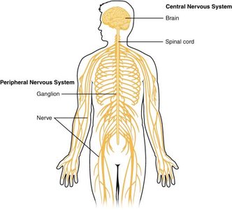
The nervous system is a complex network responsible for communication and control throughout the body. It is divided into two major regions: the Central Nervous System (CNS) and the Peripheral Nervous System (PNS).
CNS: Composed of the brain and spinal cord; responsible for processing and integrating information.
PNS: Includes all nervous tissue outside the CNS; connects the CNS to limbs and organs.
Cell Types in Nervous Tissue
Nervous tissue contains two basic types of cells:
Neurons: The communicative cells that transmit electrical signals.
Glial cells: Provide structural and metabolic support for neurons.
Anatomy of Neurons
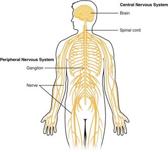
Neurons are specialized for the transmission of electrical signals. Their structure includes:
Cell body (soma): Contains the nucleus and most organelles.
Dendrites: Extensions that receive signals from other neurons.
Axon: A single, long process that transmits signals to target cells; may branch to communicate with multiple targets.
Axon terminal: Ends in synaptic end bulbs, forming synapses with target cells.
Myelin sheath: Insulating layer produced by glial cells, facilitating rapid signal transmission.
Nodes of Ranvier: Gaps in the myelin sheath important for saltatory conduction.
Organization of Nervous Tissue
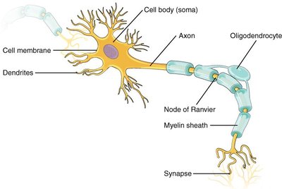
Nucleus (CNS): Localized collection of neuron cell bodies in the CNS.
Ganglion (PNS): Localized collection of neuron cell bodies in the PNS.
Tract (CNS): Bundle of axons in the CNS.
Nerve (PNS): Bundle of axons in the PNS.
Functional Divisions of the Nervous System
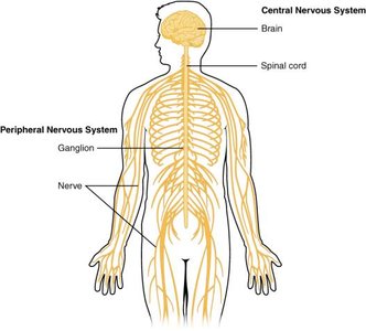
The nervous system performs three basic functions:
Sensation: Detects changes (stimuli) within the body or environment via sensory structures.
Response: Produces responses based on perceived stimuli; can be voluntary (somatic nervous system) or involuntary (autonomic nervous system).
Integration: Processes and integrates sensory information in the CNS, leading to specific responses.
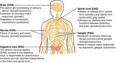
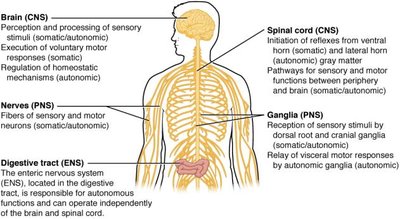
Somatic, Autonomic, and Enteric Nervous Systems
Somatic Nervous System (SNS): Controls voluntary motor responses (skeletal muscle contraction).
Autonomic Nervous System (ANS): Controls involuntary functions (smooth muscle, cardiac muscle, glands); maintains homeostasis.
Enteric Nervous System (ENS): Controls smooth muscle and glandular tissue in the digestive system; sometimes considered part of the ANS.
Types of Neurons
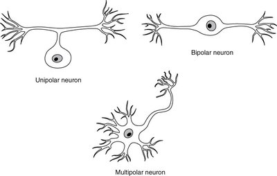
Neurons are classified based on their structure:
Pseudo-unipolar neurons: One process emerging from the cell body, which splits into two branches; sensory neurons in humans.
Bipolar neurons: Two processes (one axon, one dendrite); found in olfactory epithelium and retina.
Multipolar neurons: One axon and two or more dendrites; most common type.
Glial Cells (Neuroglia)
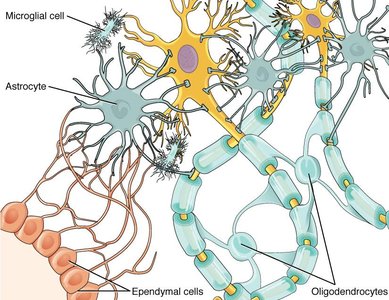
Glial cells support neurons and are classified by location:
CNS Glial Cells:
Astrocytes: Regulate ion concentrations, form blood-brain barrier, remove excess neurotransmitters.
Oligodendrocytes: Myelinate axons in the CNS.
Ependymal cells: Produce cerebrospinal fluid (CSF).
Microglial cells: Immune defense in the CNS.
PNS Glial Cells:
Satellite cells: Support neurons in ganglia, similar to astrocytes.
Schwann cells: Myelinate axons in the PNS; each wraps around one axon segment.
Membrane Potential and Ion Channels
The cell membrane separates intracellular and extracellular environments, maintaining a difference in charge known as the membrane potential.
Ion channels: Allow ions to move across the membrane; types include ligand-gated, mechanically gated, and voltage-gated channels.
Resting membrane potential: Typically, -70 mV; maintained by sodium-potassium pumps and ion leakage channels.
Types of Ion Channels
Ligand-gated: Open in response to binding of signaling molecules (ligands) such as neurotransmitters.
Mechanically gated: Open in response to physical distortion (e.g., pressure).
Voltage-gated: Open in response to changes in membrane potential.
Action Potential
An action potential is a rapid change in membrane potential that allows neurons to communicate.
Depolarization: Sodium ions enter the cell, making the inside less negative.
Threshold: If membrane potential reaches -55 mV, voltage-gated sodium channels open.
Peak: Membrane potential reaches +30 mV.
Repolarization: Potassium ions exit the cell, restoring negative charge.
Hyperpolarization: Temporary overshoot to -90 mV before returning to resting potential. `
All-or-none principle: Action potentials occur only if threshold is reached; all action potentials peak at the same voltage.
Key Equations
Resting membrane potential:
Threshold potential:
Peak action potential:
Graded Potentials
Local changes in membrane potential caused by opening of ligand-gated or mechanically gated channels.
A large enough graded potential can trigger an action potential.
Summary Table: Nervous System Components and Functions
Component | Location | Function |
|---|---|---|
Brain (CNS) | Head | Processing sensory stimuli, motor responses, homeostasis |
Spinal cord (CNS) | Vertebral column | Reflexes, sensory/motor pathways |
Nerves (PNS) | Throughout body | Transmit sensory and motor signals |
Ganglia (PNS) | Near spinal cord/limbs | Sensory reception, relay motor responses |
Digestive tract (ENS) | Digestive system | Autonomous control of digestion |
Additional info:
Action potentials are fundamental for neural communication and are the basis for all nervous system functions.
Glial cells are essential for maintaining the health and function of neurons, including myelination and immune defense.
