 Back
Chapter 13: The Nervous System – Structure, Function, and Cellular Organization
Study Guide - Smart Notes
 Tailored notes based on your materials, expanded with key definitions, examples, and context.
Nervous System: Overview and Anatomical Divisions
Introduction to the Nervous System
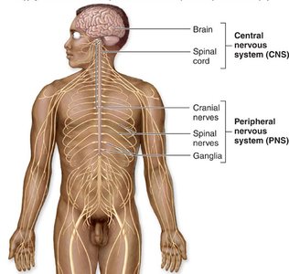
The nervous system is the primary control and communication system of the body, responsible for integrating sensory input, coordinating motor output, and regulating homeostasis. It is divided into two main anatomical divisions: the Central Nervous System (CNS) and the Peripheral Nervous System (PNS).
Central Nervous System (CNS): Consists of the brain and spinal cord; responsible for processing and integrating information.
Peripheral Nervous System (PNS): Composed of cranial nerves, spinal nerves, and ganglia; transmits information to and from the CNS.
Functional Organization of the Nervous System
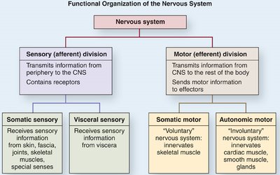
The nervous system is also organized functionally into sensory (afferent) and motor (efferent) divisions, each with somatic and visceral components.
Sensory (Afferent) Division: Transmits sensory information from receptors to the CNS.
Motor (Efferent) Division: Transmits motor commands from the CNS to effectors (muscles and glands).
Somatic: Relates to voluntary control (skeletal muscle).
Autonomic: Relates to involuntary control (cardiac muscle, smooth muscle, glands).
Neuron Structure and Function
Basic Structure of a Neuron
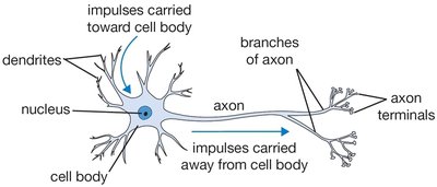
Neurons are specialized cells that conduct electrical impulses. They have a unique structure that supports their function in communication.
Cell Body (Soma): Contains the nucleus and most organelles; metabolic center of the neuron.
Dendrites: Short, branched processes that receive signals and carry them toward the cell body.
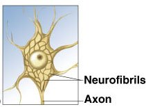
Axon: Long process that carries impulses away from the cell body to other neurons or effectors.
Axon Terminals: Branches at the end of the axon that form synapses with other cells.
Specialized Neuronal Structures
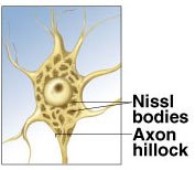
Axon Hillock: The region where the axon originates from the cell body; site of action potential initiation.
Neurofibrils: Intermediate filaments for structural support and intracellular transport.
Nissl Bodies: Clusters of rough endoplasmic reticulum and ribosomes; site of protein synthesis.
Myelin Sheath and Nodes of Ranvier
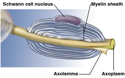
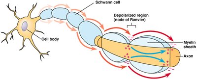
The myelin sheath is a multilayered, lipid-rich covering that insulates axons, increasing the speed of impulse conduction. Gaps in the myelin sheath, called nodes of Ranvier, facilitate saltatory conduction.
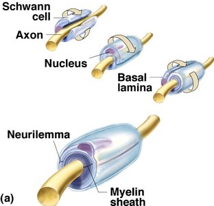
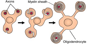
Myelin Sheath: Produced by Schwann cells in the PNS and oligodendrocytes in the CNS.
Nodes of Ranvier: Gaps between myelin segments where action potentials are regenerated.
Connective Tissue Components of Nerves
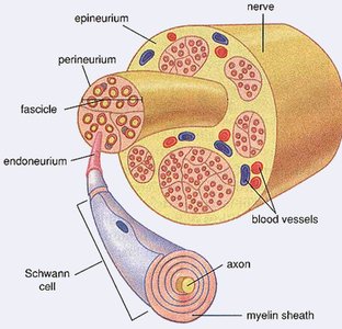
Nerves are organized into bundles and surrounded by connective tissue layers:
Epineurium: Surrounds the entire nerve.
Perineurium: Surrounds each fascicle (bundle of axons).
Endoneurium: Surrounds individual axons (external to the myelin sheath).
Classification of Neurons
Structural Classification
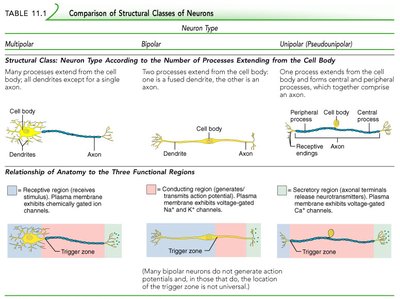
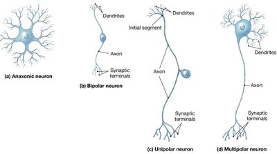
Neurons are classified by the number of processes extending from the cell body:
Type | Structure | Location/Function |
|---|---|---|
Multipolar | Many dendrites, one axon | Most common; motor neurons and interneurons in CNS |
Bipolar | One dendrite, one axon | Special senses (retina, olfactory epithelium) |
Unipolar (Pseudounipolar) | Single process splits into two branches | Sensory neurons in PNS |
Functional Classification
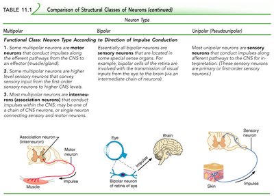
Neurons are also classified by the direction of impulse conduction:
Sensory (Afferent) Neurons: Transmit impulses from receptors to the CNS; mostly unipolar.
Motor (Efferent) Neurons: Transmit impulses from the CNS to effectors; mostly multipolar.
Interneurons (Association Neurons): Connect sensory and motor neurons within the CNS; multipolar and comprise the majority of neurons.
Neuroglia (Glial Cells)
Types and Functions of Neuroglia
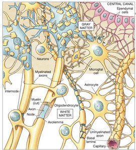
Neuroglia are supporting cells in the nervous system, outnumbering neurons by about 10:1. They provide structural support, insulation, and metabolic support for neurons.
Schwann Cells (PNS): Myelinate axons in the PNS; each cell myelinates a single axon segment.
Satellite Cells (PNS): Surround neuron cell bodies in ganglia; regulate the environment around neurons.
Oligodendrocytes (CNS): Myelinate axons in the CNS; each cell can myelinate multiple axons.
Astrocytes (CNS): Maintain the blood-brain barrier, regulate ion/nutrient concentrations, and repair injury.
Microglia (CNS): Phagocytic cells that remove debris, waste, and pathogens.
Ependymal Cells (CNS): Line brain ventricles and spinal cord central canal; produce and circulate cerebrospinal fluid (CSF).
Regeneration of Nerve Fibers
Axons in the PNS can regenerate after injury, primarily due to the activity of Schwann cells. Regeneration in the CNS is limited due to the inhibitory environment and lack of supportive glial activity.
Key Factors: Extent of damage, presence of Schwann cells, and distance between injury and effector.
Summary Table: CNS vs PNS
Feature | CNS | PNS |
|---|---|---|
Collection of Cell Bodies | Nucleus | Ganglion |
Bundle of Axons | Tract | Nerve |
Myelination by | Oligodendrocytes | Schwann Cells |
