 Back
Chapter 16: Blood – Anatomy & Physiology Study Guide
Study Guide - Smart Notes
 Tailored notes based on your materials, expanded with key definitions, examples, and context.
Blood: Internal Transport System
Introduction to Blood
Blood is a specialized connective tissue that serves as the body's internal transport system, delivering essential substances and removing waste products. It plays a critical role in maintaining homeostasis, supporting immune function, and facilitating communication between organs.
Functions of Blood:
Transport of oxygen, nutrients, hormones, and waste products
Regulation of body temperature, pH, and fluid balance
Protection against blood loss (clotting) and infection (immune response)
Composition of Blood
Major Components of Whole Blood
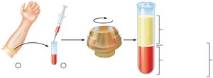
Blood consists of plasma and formed elements. Plasma is the liquid matrix, while formed elements include erythrocytes, leukocytes, and platelets.
Plasma: 55% of whole blood; least dense component
Buffy coat: Leukocytes and platelets; <1% of whole blood
Erythrocytes: 45% of whole blood (hematocrit); most dense component
Blood Plasma
Blood plasma is a straw-colored, viscous fluid composed primarily of water, dissolved solutes, and plasma proteins. It serves as the medium for transporting formed elements and dissolved substances.
Water: 90% of plasma volume; acts as solvent and temperature buffer
Solutes: Electrolytes, plasma proteins (albumin, globulins, fibrinogen), nutrients, gases, hormones, wastes
Plasma Proteins:
Albumin: Maintains osmotic pressure and transports substances
Globulins: Immune function and transport
Fibrinogen: Blood clotting
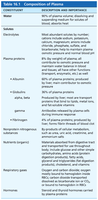
Component | Description and Importance |
|---|---|
Water | 90% of plasma volume; dissolving and suspending medium for solutes; absorbs heat |
Electrolytes | Most abundant solutes; help maintain plasma osmotic pressure and normal blood pH |
Plasma proteins | 8% of plasma; includes albumin, globulins, and fibrinogen |
Albumin | 60% of plasma proteins; maintains osmotic pressure |
Globulins | 36% of plasma proteins; transport and immune functions |
Fibrinogen | 4% of plasma proteins; forms fibrin threads in blood clotting |
Nutrients | Materials for cell metabolism |
Respiratory gases | O2 and CO2 |
Hormones | Regulatory molecules |
Formed Elements
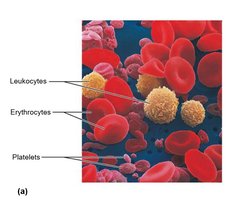
The formed elements of blood include erythrocytes (red blood cells), leukocytes (white blood cells), and platelets. These elements are suspended in plasma and perform specialized functions.
Erythrocytes: Transport oxygen and carbon dioxide
Leukocytes: Defend against infection
Platelets: Essential for blood clotting
Erythrocytes (Red Blood Cells)
Structural Characteristics of Erythrocytes
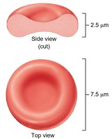
Erythrocytes are biconcave, anucleate cells optimized for gas transport. Their shape increases surface area for efficient diffusion and flexibility for passage through capillaries.
Size: Approximately 7.5 μm in diameter and 2.5 μm thick
Shape: Biconcave disc; lacks nucleus and organelles
Function of Erythrocytes
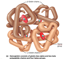
The primary function of erythrocytes is to transport oxygen from the lungs to tissues and carbon dioxide from tissues to the lungs. This is accomplished by the protein hemoglobin.
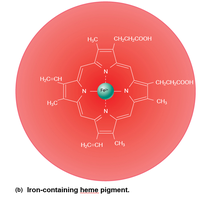
Hemoglobin: A globular protein composed of four polypeptide chains (two alpha, two beta) and four heme groups
Oxygen binding: Each heme group contains iron, which binds oxygen reversibly
Production of Erythrocytes (Erythropoiesis)
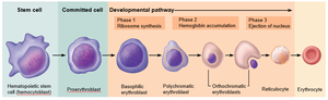
Erythropoiesis is the process of red blood cell formation, occurring primarily in the red bone marrow. It involves differentiation from hematopoietic stem cells to mature erythrocytes.
Stages: Hematopoietic stem cell → Proerythroblast → Erythroblast (various stages) → Reticulocyte → Erythrocyte
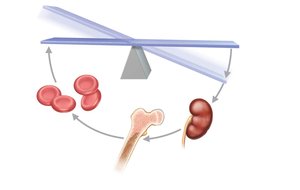
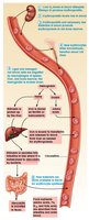
Regulation: Controlled by erythropoietin (EPO), a hormone produced by the kidneys in response to hypoxia
Regulation of Erythropoiesis
Erythropoiesis is tightly regulated to maintain adequate oxygen-carrying capacity. EPO stimulates red bone marrow to increase RBC production when oxygen levels are low.
Stimulus: Hypoxia (low O2 levels) triggers EPO release
Feedback: Increased RBCs restore oxygen levels, reducing EPO production
Clinical Note: EPO Abuse
Artificial EPO is sometimes abused by athletes to increase stamina. However, excessive RBC production can lead to dangerous consequences such as blood thickening, clotting, stroke, or heart failure.
Fate and Destruction of Erythrocytes
Erythrocytes have a lifespan of about 120 days. Old or damaged RBCs are removed by macrophages in the spleen, liver, and bone marrow. Hemoglobin is broken down into heme and globin; heme is further degraded to bilirubin and excreted.
Erythrocyte Disorders
Anemia
Anemia is a condition characterized by insufficient RBCs or hemoglobin, resulting in reduced oxygen delivery to tissues. Causes include blood loss, decreased RBC production, or increased RBC destruction.
Sickle-Cell Anemia
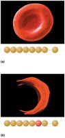
Sickle-cell anemia is a genetic disorder caused by a single amino acid substitution in the beta chain of hemoglobin. This results in abnormal, sickle-shaped erythrocytes that can block blood vessels and cause pain.
Normal hemoglobin: Glu at position 6
Sickled hemoglobin: Val at position 6
Leukocytes (White Blood Cells)
General Structure and Functional Characteristics
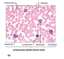
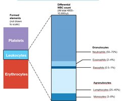
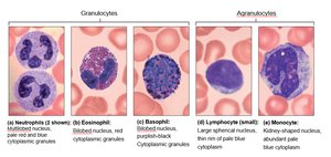
Leukocytes are nucleated cells that defend the body against infection and foreign substances. They are classified as granulocytes or agranulocytes based on the presence of cytoplasmic granules.
Granulocytes: Neutrophils, eosinophils, basophils
Agranulocytes: Lymphocytes, monocytes
Granulocytes Overview
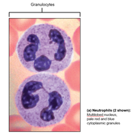
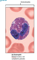
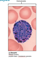
Granulocytes contain visible granules in their cytoplasm and have lobed nuclei. They are involved in immune responses and inflammation.
Neutrophils: Phagocytize bacteria; most abundant
Eosinophils: Combat parasitic infections and modulate allergic responses
Basophils: Release histamine; involved in inflammatory responses
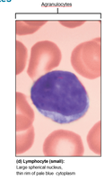
Agranulocytes Overview
Agranulocytes lack visible granules and have simpler nuclei. They play roles in adaptive immunity and phagocytosis.
Lymphocytes: B and T cells; responsible for specific immune responses
Monocytes: Differentiate into macrophages; phagocytize pathogens and debris
Leukopoiesis: Formation of WBCs
Leukopoiesis
Leukopoiesis is the process of white blood cell formation, originating from hematopoietic stem cells in the bone marrow. It is regulated by cytokines and growth factors.
Myeloid stem cells: Give rise to granulocytes and monocytes
Lymphoid stem cells: Give rise to lymphocytes
Clinical Note: Hematopoietic Hormones
Hematopoietic hormones such as EPO and colony-stimulating factors (CSFs) are used clinically to stimulate bone marrow in patients undergoing chemotherapy or with immune deficiencies.
Platelets
Structure, Function, and Formation
Platelets are cell fragments derived from megakaryocytes. They are essential for blood clotting and repair of damaged blood vessels.
Formation: Megakaryocytes fragment to release platelets into circulation
Function: Form platelet plugs and release factors for coagulation
Hemostasis
Events of Hemostasis
Hemostasis is the process that stops bleeding through a series of steps: vascular spasm, platelet plug formation, and coagulation.
Vascular spasm: Smooth muscle contracts to reduce blood flow
Platelet plug formation: Platelets adhere to exposed collagen and aggregate
Coagulation: Fibrin forms a mesh that stabilizes the clot
Blood Groups, Transfusions, and Diagnostic Tests
Transfusing Red Blood Cells: Antigens and Blood Groups
Blood groups are determined by the presence of antigens on the surface of erythrocytes. The ABO and Rh systems are clinically significant for transfusions.
ABO system: Type A, B, AB, O based on antigen presence
Rh system: Rh+ or Rh– based on D antigen
Clinical Note: Hemolytic Disease of the Newborn
Hemolytic disease of the newborn occurs when an Rh– mother develops antibodies against Rh+ fetal blood, leading to destruction of fetal RBCs in subsequent pregnancies.
Blood Typing and Cross Matching
Blood typing and cross matching are essential for safe transfusions, ensuring compatibility between donor and recipient blood.
Key Takeaways
Blood is a vital connective tissue with transport, regulatory, and protective functions
Plasma and formed elements are the main components
Erythrocytes, leukocytes, and platelets each have specialized roles
Hemostasis prevents blood loss and maintains vascular integrity
Blood groups are critical for transfusion safety
