 Back
BackChapter 16: Blood – Structure, Function, and Physiology
Study Guide - Smart Notes
 Tailored notes based on your materials, expanded with key definitions, examples, and context.
Blood: Structure, Function, and Physiology
Overview
Blood is a specialized connective tissue essential for the transport of gases, nutrients, waste products, and immune cells throughout the body. It consists of a fluid matrix called plasma and various cellular elements, each with distinct physiological roles.
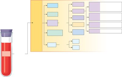
Plasma and the Cellular Elements of Blood
Plasma Composition
Plasma is the extracellular matrix of blood, making up about 55% of total blood volume. It is similar to interstitial fluid but contains unique plasma proteins.
Water (~92%): Solvent for carrying other substances.
Proteins (~7%): Includes albumins, globulins, fibrinogen, and transferrin.
Other Solutes (~1%): Ions (Na+, K+, Cl-, HCO3-), organic molecules (glucose, lipids, amino acids), gases (O2, CO2), trace elements, and vitamins.
Plasma Proteins:
Albumins: Major contributors to colloid osmotic pressure; carriers for various substances.
Globulins: Clotting factors, enzymes, antibodies, and carriers.
Fibrinogen: Forms fibrin threads essential for blood clotting.
Transferrin: Iron transport.
Cellular Elements of Blood
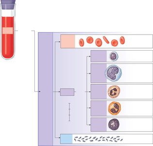
The cellular elements include red blood cells (RBCs), white blood cells (WBCs), and platelets.
Red Blood Cells (Erythrocytes): Transport oxygen and carbon dioxide.
White Blood Cells (Leukocytes): Immune defense; subdivided into:
Lymphocytes (Immunocytes): Specific immune responses.
Monocytes: Develop into macrophages; phagocytic.
Neutrophils: Phagocytes and granulocytes.
Eosinophils: Granulocytes; combat parasites and participate in allergic responses.
Basophils: Granulocytes; tissue basophils are called mast cells.
Platelets: Cell fragments essential for blood clotting.
Blood Cell Production (Hematopoiesis)
Sites and Regulation of Hematopoiesis
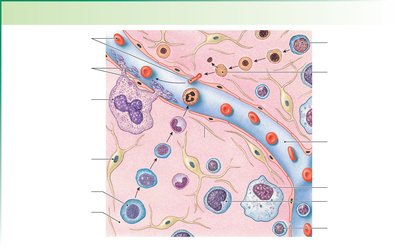
Blood cells are produced in the bone marrow through a process called hematopoiesis. Red bone marrow is active and contains hemoglobin, while yellow bone marrow is inactive and stores fat.
Hematopoiesis: Production of all blood cells.
Leukopoiesis: Production of white blood cells.
Erythropoiesis: Production of red blood cells.
Thrombopoiesis: Production of platelets.
Regulatory Cytokines:
Colony Stimulating Factors (CSFs): Regulate leukopoiesis; produced by bone marrow cells.
Thrombopoietin (TPO): Regulates platelet production; produced in the liver.
Erythropoietin (EPO): Regulates red blood cell production; produced in the kidney.
Red Blood Cells (Erythrocytes)
Structure and Function
Red blood cells are specialized for oxygen transport. They lack a nucleus at maturity and have a biconcave disk shape, which increases surface area for gas exchange.
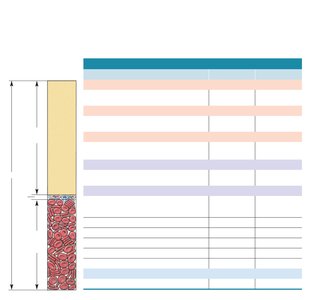
Hematocrit: Ratio of RBCs to plasma, expressed as a percentage.
Mean Corpuscular Volume (MCV): Average volume of one RBC.
Mean Corpuscular Hemoglobin (MCH): Amount of hemoglobin per RBC.
Mean Corpuscular Hemoglobin Concentration (MCHC): Hemoglobin per volume of RBC.

Osmotic Effects and Abnormal Morphology
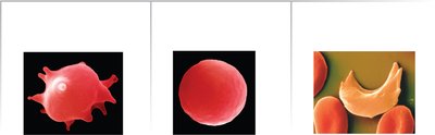
In hypertonic solutions, RBCs shrink and become crenated.
In hypotonic solutions, RBCs swell and lose their shape.
Sickle cell disease causes abnormal hemoglobin, leading to sickled RBCs.
Hemoglobin Synthesis and Iron Metabolism
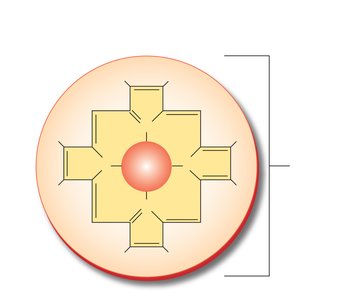
Hemoglobin is a protein with four globin chains, each containing a heme group with an iron atom. Iron is essential for hemoglobin synthesis and is transported by transferrin and stored as ferritin in the liver.
Hemoglobin: Four polypeptide chains (2 alpha, 2 beta) and four heme groups.
Iron: Obtained from diet, transported by transferrin, stored as ferritin.
Red Blood Cell Life Cycle and Disorders
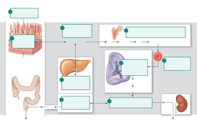
RBCs live about 120 days; old cells are destroyed in the spleen.
Hemoglobin breakdown produces bilirubin, excreted in bile and urine.
Disorders include anemia (low RBCs or hemoglobin), hemolytic anemia, sickle cell disease, and iron-deficiency anemia.
Jaundice results from elevated bilirubin levels.
Platelets
Structure and Function
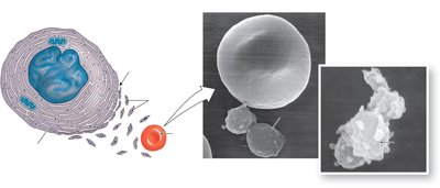
Platelets are small cell fragments derived from megakaryocytes. They are essential for blood clotting, immunity, and inflammation, with a lifespan of about 10 days.
Contain vesicles with cytokines and growth factors.
Activated platelets become spiky and adhere to each other.
Hemostasis and Coagulation
Hemostasis: Prevention of Blood Loss
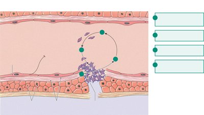
Hemostasis is the process that prevents blood loss from damaged vessels and involves three major steps:
Vasoconstriction: Narrowing of blood vessels due to paracrines from endothelium and platelets.
Platelet Plug Formation: Platelets adhere to exposed collagen, become activated, and aggregate to form a plug.
Coagulation: Formation of a fibrin clot to reinforce the platelet plug.
Coagulation Cascade and Fibrinolysis
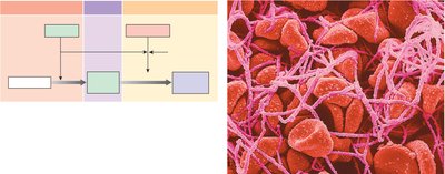
Coagulation involves a cascade of enzymatic reactions leading to the conversion of fibrinogen to fibrin, forming a stable clot. There are intrinsic and extrinsic pathways, both converging to activate thrombin. Fibrinolysis is the breakdown of the clot as the vessel heals.
Intrinsic Pathway: Triggered by contact activation.
Extrinsic Pathway: Triggered by tissue injury and tissue factor.
Common Pathway: Both pathways lead to thrombin formation, which converts fibrinogen to fibrin.
Fibrinolysis: Thrombin and tissue plasminogen activator (tPA) convert plasminogen to plasmin, which digests fibrin.

Hemophilia
Hemophilia is a group of genetic disorders where one or more factors in the coagulation cascade are defective or missing. Hemophilia A is the most common form and primarily affects males, leading to uncontrolled bleeding.
Summary Table: Complete Blood Count (CBC) Normal Ranges
Test | Males | Females |
|---|---|---|
Hematocrit (%) | 40–54 | 37–47 |
Hemoglobin (g/dL) | 14–17 | 12–16 |
Red Cell Count (cells/µL) | 4.5–6.5 × 10⁶ | 3.9–5.6 × 10⁶ |
Total White Count (cells/µL) | 4–11 × 103 | 4–11 × 103 |
Neutrophils (%) | 50–70 | 50–70 |
Eosinophils (%) | 1–4 | 1–4 |
Basophils (%) | <1 | <1 |
Lymphocytes (%) | 20–40 | 20–40 |
Monocytes (%) | 2–8 | 2–8 |
Platelets (per µL) | 150–450 × 103 | 150–450 × 103 |
Key Equations
Hematocrit (%):
