 Back
BackChapter 16: Blood – Structure, Function, and Clinical Applications
Study Guide - Smart Notes
 Tailored notes based on your materials, expanded with key definitions, examples, and context.
Blood: Composition and Functions
Plasma and Cellular Elements
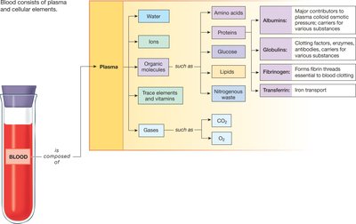
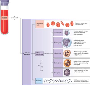
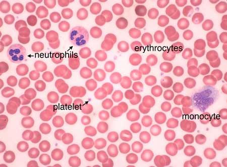
Blood is a specialized connective tissue composed of plasma (the fluid matrix) and cellular elements. Plasma constitutes about 58% of blood volume and contains water, proteins, ions, organic molecules, trace elements, vitamins, and gases. The cellular elements include red blood cells (RBCs), white blood cells (WBCs), and platelets.
Plasma: Contains water (~92%), proteins (~7%), and other solutes (~1%).
Plasma Proteins: Albumins (osmotic pressure, carriers), globulins (clotting, defense), fibrinogen (clotting), transferrin (iron transport).
Cellular Elements: RBCs (oxygen and carbon dioxide transport), WBCs (immune defense), platelets (clotting).
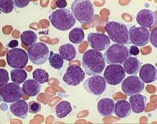
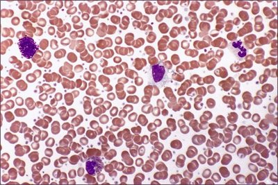
White Blood Cells (Leukocytes)
Types and Functions
White blood cells are the least abundant formed elements in blood, but are essential for immune defense. They are classified based on the presence of granules:
Agranulocytes: Monocytes (phagocytic, become macrophages), lymphocytes (antibody production, immune memory).
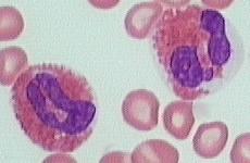
Granulocytes: Neutrophils (phagocytic, most abundant), eosinophils (defense against parasites, allergies), basophils (release histamine and heparin).
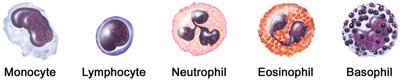
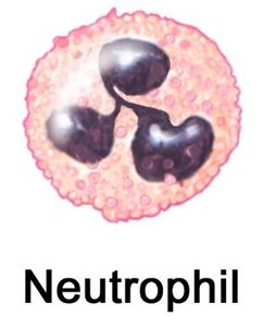
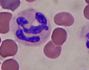
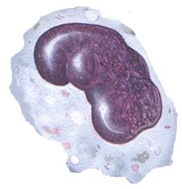
Neutrophils
Neutrophils are the most abundant WBCs (60-70%), with a multi-lobed nucleus. They respond rapidly to bacterial infections and are key phagocytes.
Polymorphonuclear leukocytes (PMNs): Another name for neutrophils due to their lobed nuclei.
Band cells: Immature neutrophils.
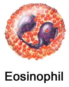
Eosinophils
Eosinophils are less common (2-4%), with large red-orange granules. They are abundant in mucous membranes and increase in allergies and parasitic infections.
Eosinophilia: Elevated eosinophil count in response to certain diseases.
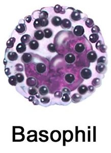
Basophils
Basophils are the rarest WBCs (<0.5%), with dark violet granules. They release histamine (vasodilation) and heparin (anticoagulant), attracting other WBCs to sites of infection.
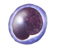
Lymphocytes
Lymphocytes are agranulocytes, making up 25-33% of WBCs. They have a round nucleus and are responsible for antibody production, immune memory, and antigen presentation.
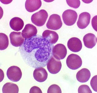
Monocytes
Monocytes are the largest WBCs (3-8%), with a kidney-shaped nucleus. They become macrophages in tissues and are highly phagocytic and antigen-presenting.
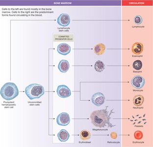
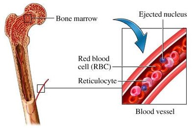
Blood Cell Production (Hematopoiesis)
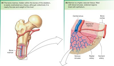
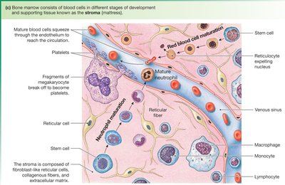
Bone Marrow and Stem Cells
Blood cells are produced in the bone marrow through hematopoiesis, regulated by cytokines. Red bone marrow is active and contains hemoglobin, while yellow marrow is inactive and stores fat.
Pluripotent hematopoietic stem cells: Can differentiate into any blood cell type.
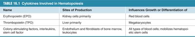
Cytokines: Interleukins, colony-stimulating factors (CSFs), erythropoietin (EPO), thrombopoietin (TPO).
Cytokines Involved in Hematopoiesis
Cytokines regulate the growth and differentiation of blood cells. The main cytokines are:
Name | Sites of Production | Influences Growth or Differentiation of |
|---|---|---|
Erythropoietin (EPO) | Kidney cells primarily | Red blood cells |
Thrombopoietin (TPO) | Liver primarily | Megakaryocytes |
Colony-stimulating factors, interleukins, stem cell factor | Endothelium and fibroblasts of bone marrow, leukocytes | All types of blood cells; mobilizes hematopoietic stem cells |
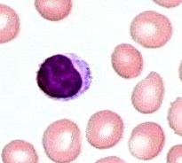
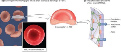
Red Blood Cells (Erythrocytes)
Structure and Function
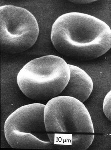
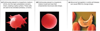
Red blood cells are biconcave, lack a nucleus when mature, and are specialized for oxygen and carbon dioxide transport. Their morphology can indicate disease.
Hematocrit: Ratio of RBCs to plasma, expressed as a percentage.
Mean corpuscular volume (MCV): Average size of RBCs.
Clinical Applications: Reticulocytes and RBC Disorders
Reticulocytes are immature RBCs, accounting for 1-2% of circulating erythrocytes. Their count indicates the rate of RBC formation. Disorders include anemia (too few RBCs), polycythemia (too many RBCs), and sickle cell disease (abnormal hemoglobin).
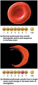
Sickle cell disease: Caused by a single amino acid change in hemoglobin, leading to crescent-shaped RBCs and poor oxygen delivery.
Anemia: Can result from inadequate erythropoiesis, hemorrhage, or hemolysis.
Polycythemia: Excess RBCs, increasing blood viscosity and risk of embolism.
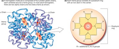
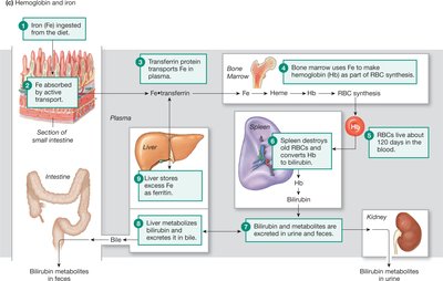
Hemoglobin and Iron Metabolism
Hemoglobin Structure and Function
Hemoglobin is a protein composed of four globin chains, each with a heme group containing iron. It is essential for oxygen transport.
Heme: Porphyrin ring with an iron atom at its center.
Iron metabolism: Iron is absorbed from the diet, transported by transferrin, stored in the liver by ferritin, and used in bone marrow for hemoglobin synthesis.
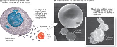
Platelets and Hemostasis
Platelet Structure and Function
Platelets are cell fragments derived from megakaryocytes, essential for blood clotting, immunity, and inflammation. They have a 10-day lifespan and contain granules with cytokines and growth factors.
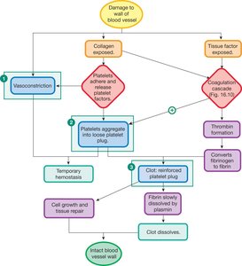
Hemostasis and Coagulation
Hemostasis prevents blood loss from damaged vessels through vasoconstriction, platelet plug formation, and coagulation. The coagulation cascade involves intrinsic and extrinsic pathways, converging to form a fibrin clot.
Intrinsic pathway: Activated by exposed collagen.
Extrinsic pathway: Activated by tissue factor.
Common pathway: Thrombin converts fibrinogen to fibrin.
Fibrinolysis: Plasmin breaks down fibrin, dissolving the clot.
Anticoagulants and Hemophilia
Endogenous anticoagulants (heparin, antithrombin III, protein C) limit clotting. Hemophilia is caused by defects in coagulation factors, leading to impaired clot formation.
Factors Involved in Coagulation
Coagulation involves multiple chemical factors, each with specific roles:
Chemical Factor | Source | Activated by/Released in Response to | Role in Coagulation | Other Roles/Comments |
|---|---|---|---|---|
Collagen | Subendothelial extracellular matrix | Injury exposing collagen | Starts intrinsic pathway | N/A |
von Willebrand factor (vWF) | Endothelium, megakaryocytes | Exposure to collagen | Regulates factor VIII | Deficiency causes prolonged bleeding |
Kininogen and kallikrein | Liver and plasma | Cofactors in plasma pathway | Contact activation of intrinsic pathway | Mediate inflammation, enhance fibrinolysis |
Tissue factor (factor III) | Most cells except platelets | Damage to tissue | Starts extrinsic pathway | N/A |
Prothrombin and thrombin (factor II) | Liver and plasma | Platelet lipids, factor V | Fibrin production | N/A |
Fibrinogen and fibrin (factor I) | Liver and plasma | Thrombin | Form insoluble fibers | N/A |
Fibrin-stabilizing factor (XIII) | Liver, megakaryocytes | Platelets | Cross-links fibrin polymers | N/A |
Ca2+ (factor IV) | Plasma ions | N/A | Required for several steps | Never a limiting factor |
Vitamin K | Diet | N/A | Synthesis of factors II, VII, IX, X | N/A |
Key Equations and Definitions
Hematocrit (%):
Mean Corpuscular Volume (MCV):
Hemoglobin Structure:
Summary Table: Normal Blood Values
Test | Males | Females |
|---|---|---|
Hematocrit (%) | 40–54 | 37–47 |
Hemoglobin (g/dL) | 14–17 | 12–16 |
Red Cell Count (cells/mL) | 4.5–6.5×10⁶ | 3.9–5.6×10⁶ |
Total White Count (cells/mL) | 4–11×103 | 4–11×103 |
Platelets (per mL) | 150–450×103 | 150–450×103 |
Clinical Applications
Leukopenia: Low WBC count (<5,000/µL), often due to toxins, infections, or drugs.
Leukocytosis: High WBC count (>10,000/µL), often due to infection, allergy, or stress.
Leukemia: Cancer of hemopoietic tissues, producing excessive immature WBCs.
Anemia: Reduced RBCs or hemoglobin, leading to decreased oxygen transport.
Polycythemia: Excess RBCs, increasing risk of embolism and cardiovascular complications.
Hemophilia: Deficiency in coagulation factors, causing prolonged bleeding.
